Category:ECG
Idi na navigaciju
Idi na pretragu
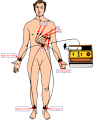
method to record the electrical activity of the heart through passive electrodes placed over the skin.  | |||||
| Postavi datoteku | |||||
| Je |
| ||||
|---|---|---|---|---|---|
| Je podklasa od |
| ||||
| Je dio | |||||
| |||||
Potkategorije
Prikazano je 13 potkategorija, od ukupno 13.
- ECG on stamps (3 F)
- Videos of ECG (5 F)
B
- Benign early repolarization (2 F)
D
- Doing ECG (22 F)
E
G
H
I
M
- Media from CardioNetworks ECGpedia (4603 F)
P
S
- Stress echocardiography (35 F)
Datoteke u kategoriji "ECG"
Ova kategorija ima slijedeću 71 datoteku.
-
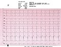
 12 lead ECG.jpg 3.507 × 2.300; 1,59 MB
-
 1秒的心电图纸及举例和注释.svg 480 × 350; 29 KB
-
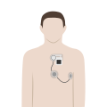
 202305 Electrocardiogram Child.svg 512 × 512; 280 KB
-
 202305 Electrocardiogram Female.svg 512 × 512; 282 KB
-
 202305 Electrocardiogram Male.svg 512 × 512; 283 KB
-
 202310 Holter Electrocardiography.svg 512 × 512; 157 KB
-
 202310 Quantitative Electroencephalography Child.svg 512 × 512; 520 KB
-
 202310 Quantitative Electroencephalography Female.svg 512 × 512; 511 KB
-
 202310 Quantitative Electroencephalography Male.svg 512 × 512; 497 KB
-
 24-hour pH-metry and electrocardiography (Gastroscan-ECG).jpg 1.056 × 2.073; 598 KB
-
 American practitioner (1912) (14597420440).jpg 2.816 × 1.618; 653 KB
-
 Annotated ECGcolor.svg 554 × 714; 378 KB
-
 Aort insuf.jpg 531 × 424; 50 KB
-
 Archery nobel prize, Museum Boerhaave Leiden.jpg 3.456 × 5.184; 5,08 MB
-
 AVD principle.jpg 600 × 288; 44 KB
-
 Avd prinzip.jpg 643 × 285; 51 KB
-

-

-
 Blackberry 9550でのMFERアプリ(DaSH MFER).JPG 1.632 × 1.224; 258 KB
-
 Both ECG.jpg 1.280 × 960; 253 KB
-
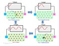
 Cells in rest en (CardioNetworks ECGpedia).png 800 × 482; 39 KB
-
 Changing ST (CardioNetworks ECGpedia).svg 667 × 479; 52 KB
-
 De-Wpw2 (CardioNetworks ECGpedia).png 800 × 600; 169 KB
-
 DRJ case 2 2 (CardioNetworks ECGpedia).jpg 711 × 617; 17 KB
-
 DRJ case 2 3 (CardioNetworks ECGpedia).jpg 711 × 617; 21 KB
-
 DVA1738 (CardioNetworks ECGpedia).jpg 619 × 511; 136 KB
-
 DVA1739 (CardioNetworks ECGpedia).jpg 474 × 391; 141 KB
-
 ECG advertising AO AL.jpg 837 × 803; 369 KB
-
 ECG processing.png 2.292 × 1.008; 252 KB
-
 ECG Ruler.png 3.065 × 856; 1,12 MB
-
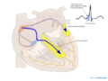
 ECG Segments Corresponding to Anatomical Structures of the Heart.jpg 2.000 × 1.027; 157 KB
-
 ECG-atrial flutter.jpg 1.730 × 1.016; 1,1 MB
-
 Eigenfrequenzen des Myokards.svg 647 × 402; 389 KB
-
 EKG of a 25 year old European male at 105 BPM.jpg 4.500 × 3.470; 6,09 MB
-
 EKG of a 27 year old European male at 78 BPM.jpg 4.499 × 3.500; 8,91 MB
-
 Ekg Schema.svg 1.085 × 1.016; 152 KB
-
EKG Sound Alert.wav 1 min 0 s; 10,02 MB
-
 Electromyogram Illustration.png 640 × 480; 281 KB
-

-
-
 Fig 7 .jpg 498 × 536; 45 KB
-
 Fig. 6..jpg 645 × 520; 58 KB
-
 Fixed LBBB (CardioNetworks ECGpedia).svg 397 × 307; 122 KB
-
 Flipped Posterior STEMI ECG.png 1.711 × 1.689; 4,41 MB
-
 G3260 Mac.png 466 × 405; 139 KB
-
 Heart transplant ECG Final.jpg 11.195 × 6.473; 2,71 MB
-
 Hopital de LaSalle - 09.jpg 1.845 × 1.385; 355 KB
-
 ICS3000 PK199.jpg 2.304 × 1.192; 1,03 MB
-
 Ion currents en (CardioNetworks ECGpedia).png 519 × 800; 138 KB
-
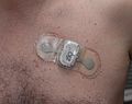 IRhythm ZIO® XT Patch.jpg 2.500 × 1.982; 670 KB
-
 Journal.pone.0141573.g004.png 1.251 × 1.720; 1,56 MB
-
 Electrocardiography; T. Lewis, 1913 Wellcome L0006119.jpg 4.480 × 3.872; 6,77 MB
-
 Localisation of the occlusion in STEMI.svg 716 × 354; 450 KB
-
 LVH with LAH.jpg 1.813 × 1.080; 1,34 MB
-
 MI colours en (CardioNetworks ECGpedia).png 800 × 494; 23 KB
-
 Mirule (CardioNetworks ECGpedia).png 800 × 626; 258 KB
-
 OpenBCI Screenshot of a Basic EKG.png 1.366 × 746; 55 KB
-
 PericarditisMyocarditis.jpg 4.204 × 2.001; 3,51 MB
-
 PLAX Mmode (CardioNetworks ECHOpedia).jpg 636 × 434; 91 KB
-
 Process of simultaneously recording ECG and SCG data.jpg 4.624 × 3.472; 4,87 MB
-
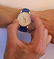 Recording single-lead ECG using a Withings Move ECG watch.jpg 1.471 × 1.591; 367 KB
-
 Right Bundle branch block.jpg 1.972 × 1.654; 707 KB
-
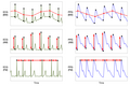
 RSA neonatal ECG+RESP+HR.png 19.200 × 6.150; 1,21 MB
-
 Sample Biosignals (ECG, PPG, RIP).svg 512 × 293; 32 KB
-
 Storia ECG.jpg 960 × 720; 72 KB
-
 Test2 (CardioNetworks ECGpedia).svg 765 × 990; 26 KB
-
 Thomas Lewis’ electrocardiograph, Cambridge, England Wellcome L0057898.jpg 3.504 × 2.735; 1,31 MB
-
 Thomas Lewis’ electrocardiograph, Cambridge, England Wellcome L0057899.jpg 3.504 × 2.725; 1,38 MB
-
 Triggering a single-lead ECG recording on a Withings Move ECG watch.jpg 1.249 × 1.544; 319 KB
-
 VolumeConductorprincipleITA.jpg 1.536 × 1.187; 186 KB
-
 دورة ECG 1.JPG 3.264 × 2.448; 2,23 MB


.jpg)
_(14597420440).jpg)


_-_DPLA_-_56ac0ed8aeb994b3196758335079e475.jpg)

.JPG)

.png)
.svg)
.png)
.jpg)
.jpg)
.jpg)
.jpg)


_and_photoplethysmogram_(PPG).png)


.svg)


.png)


.png)
.png)


.jpg)


.svg)

.svg)


