Category:ECG principle
Vai alla navigazione
Vai alla ricerca
valore pressorio rilevato nel tratto terminale della vena cava superiore e corrispondente alla pressione vigente nell'atrio destro. | |||||
| Carica un file multimediale | |||||
| Istanza di |
| ||||
|---|---|---|---|---|---|
| Sottoclasse di |
| ||||
| |||||
Sottocategorie
Questa categoria contiene le 4 sottocategorie indicate di seguito, su un totale di 4.
E
P
W
File nella categoria "ECG principle"
Questa categoria contiene 80 file, indicati di seguito, su un totale di 80.
-
 2022 Electrocardiogram.jpg 1 658 × 1 086; 566 KB
-
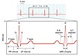
 2028 Cardiac Cycle vs Electrocardiogram.jpg 1 683 × 800; 237 KB
-
 A. Waller, Introductory address on the elect Wellcome L0031415.jpg 1 208 × 1 550; 1,02 MB
-
 A. Waller, Introductory address on the elect Wellcome L0031416.jpg 1 216 × 1 578; 915 KB
-

-
 Animation of ECG Limb Leads.gif 1 910 × 1 052; 449 KB
-
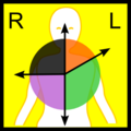 Axis deviation (ECG).png 300 × 300; 17 KB
-
 Blausen 0339 Electrocardiogram.png 640 × 480; 901 KB
-
 Blausen 0465 Heartbeat EKG.png 750 × 1 500; 100 KB
-
 Blokové schéma EGG nahrávání a analýzy.png 574 × 295; 9 KB
-
 Cabrerakreis zh-hans.png 392 × 387; 5 KB
-
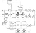 Celkové schema ekg přístroje.jpg 778 × 718; 57 KB
-
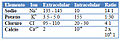 Concentraciones.jpg 326 × 111; 52 KB
-
 De-Actionpotential (CardioNetworks ECGpedia).png 800 × 600; 27 KB
-
 De-CableReversal1 (CardioNetworks ECGpedia).png 800 × 290; 22 KB
-
 De-Conduction ap (CardioNetworks ECGpedia).png 800 × 600; 146 KB
-
 De-ECG lead angulation (CardioNetworks ECGpedia).png 800 × 600; 56 KB
-
 De-ECGafleidingen (CardioNetworks ECGpedia).jpg 486 × 599; 31 KB
-
 De-Formule QTc (CardioNetworks ECGpedia).png 151 × 36; 754 byte
-
 De-Hart axis (CardioNetworks ECGpedia).png 637 × 355; 102 KB
-
 De-Hartas2 (CardioNetworks ECGpedia).jpg 605 × 600; 21 KB
-
 De-Left axis dev (CardioNetworks ECGpedia).jpg 584 × 599; 21 KB
-
 De-Milstein algorythm (CardioNetworks ECGpedia).png 800 × 591; 59 KB
-
 De-Ontstaan LBTB (CardioNetworks ECGpedia).png 409 × 262; 8 KB
-
 De-P wave morphology (CardioNetworks ECGpedia).png 800 × 600; 38 KB
-
 De-Pta changes (CardioNetworks ECGpedia).png 800 × 600; 80 KB
-
 De-Re entry (CardioNetworks ECGpedia).png 800 × 600; 50 KB
-
 De-Reduced rwaveprogression (CardioNetworks ECGpedia).png 800 × 600; 120 KB
-
 De-Rhythm LBTBmorph nl (CardioNetworks ECGpedia).png 737 × 600; 85 KB
-
 De-Rhythm RBTBmorph nl (CardioNetworks ECGpedia).png 800 × 415; 16 KB
-
 De-Rhythm RSratio (CardioNetworks ECGpedia).png 674 × 600; 32 KB
-
 De-Right axis dev (CardioNetworks ECGpedia).jpg 597 × 600; 21 KB
-
 De-Rwaveprogression (CardioNetworks ECGpedia).png 800 × 600; 117 KB
-
 De-Scn5a (CardioNetworks ECGpedia).jpg 513 × 181; 17 KB
-
 De-Stroomgebieden (CardioNetworks ECGpedia).png 747 × 599; 197 KB
-
 De-SVT overview.svg (CardioNetworks ECGpedia).png 800 × 600; 89 KB
-
 De-T wave morphology (CardioNetworks ECGpedia).png 800 × 600; 56 KB
-
 Ecg einthoven.png 600 × 598; 12 KB
-
 ECG leads - angles.png 300 × 300; 30 KB
-
 ECG leads - arrows (no names).png 300 × 300; 23 KB
-
 ECG leads - named (no arrows).png 300 × 300; 25 KB
-
 ECG Paper.jpg 480 × 360; 85 KB
-
 ECG Principle fast.gif 700 × 800; 667 KB
-
 ECG principle slow.gif 700 × 800; 671 KB
-
 ECG rS nomenclature.png 105 × 160; 3 KB
-
 ECG-Einthoven-vect6-sklon osy45.png 1 938 × 1 595; 137 KB
-
 Einthoven triangle and cardiac vector.JPG 363 × 314; 12 KB
-
 EKG leads.png 814 × 726; 381 KB
-
 Electrocardiogram demonstration Wellcome L0009603.jpg 1 440 × 1 380; 714 KB
-
 Electrocardiogram Depolarization.jpg 800 × 524; 87 KB
-
 Extreme axis deviation (ECG).png 300 × 300; 13 KB
-
 Interactions within Excitation-contraction Coupling.jpg 611 × 639; 138 KB
-
 Lead aVF (ECG).png 300 × 300; 14 KB
-
 Lead aVL (ECG).png 300 × 300; 16 KB
-
 Lead aVR (ECG).png 300 × 300; 16 KB
-
 Lead direction.png 600 × 450; 484 KB
-
 Lead I (ECG).png 300 × 300; 12 KB
-
 Lead II (ECG).png 300 × 300; 16 KB
-
 Lead III (ECG).png 300 × 300; 16 KB
-
 Left axis deviation (ECG).png 300 × 300; 14 KB
-
 Limb leads of EKG.png 1 500 × 1 000; 372 KB
-
 Mappaecg1A.JPG 525 × 374; 174 KB
-
 Normal cardiac axis (ECG).png 300 × 300; 14 KB
-
 Normal EKG axis.png 814 × 726; 435 KB
-
 Porder (ECG).png 400 × 200; 10 KB
-
 PQRS origin en (CardioNetworks ECGpedia).png 800 × 883; 287 KB
-
 QCO waves.png 267 × 188; 12 KB
-
 QRSwaves (CardioNetworks ECGpedia).jpg 800 × 820; 42 KB
-
 QTeyeballing (CardioNetworks ECGpedia).jpg 880 × 237; 27 KB
-
 QTeyeballing (CardioNetworks ECGpedia).png 880 × 237; 70 KB
-
 QTeyeballing2 (CardioNetworks ECGpedia).png 880 × 237; 70 KB
-
 Rapid Axis.jpg 720 × 289; 46 KB
-
 Right axis dev (CardioNetworks ECGpedia).jpg 800 × 804; 40 KB
-
 Right axis deviation (ECG).png 300 × 300; 14 KB
-
 Schema ekg.jpg 708 × 765; 42 KB
-
 Shortcut to ECG.pdf 725 × 1 091, 53 pagine; 2,8 MB
-
 Thoracic lead.jpg 466 × 626; 171 KB
-
 Triangolo di Einthoven.jpg 470 × 300; 59 KB
-
 Wilsonova síť.jpg 439 × 386; 14 KB
-
 Моделирование волны U.JPG 546 × 175; 21 KB


.png)


.png)
.png)
.png)
.jpg)
.png)
.jpg)
.jpg)
.png)
.png)
.png)
.png)
.png)
.png)
.png)
.png)
.png)
.jpg)
.png)
.png)
.png)
.png)


.png)
.png)


.png)

.png)
.png)
.png)

.png)
.png)
.png)
.png)


.png)

.png)
.png)

.jpg)
.jpg)
.png)


.png)
.png)
.jpg)
.jpg)
.png)
.png)