Category:Endoplasmic reticulum
Zur Navigation springen
Zur Suche springen
Zellorganell  | |||||
| Medium hochladen | |||||
| Ist ein(e) |
| ||||
|---|---|---|---|---|---|
| Unterklasse von |
| ||||
| Ist Teil von | |||||
| Physisch verbunden mit |
| ||||
| |||||
Unterkategorien
Es werden 4 von insgesamt 4 Unterkategorien in dieser Kategorie angezeigt:
In Klammern die Anzahl der enthaltenen Kategorien (K), Seiten (S), Dateien (D)
Medien in der Kategorie „Endoplasmic reticulum“
Folgende 200 Dateien sind in dieser Kategorie, von 201 insgesamt.
(vorherige Seite) (nächste Seite)-
 0313 Endoplasmic Reticulum a en.png 604 × 362; 230 KB
-
 0313 Endoplasmic Reticulum b en.png 467 × 363; 190 KB
-
 0313 Endoplasmic Reticulum b labeled.png 371 × 363; 196 KB
-
 0313 Endoplasmic Reticulum c en.png 733 × 398; 245 KB
-
 0313 Endoplasmic Reticulum c labeled.png 604 × 398; 250 KB
-
 0313 Endoplasmic Reticulum.jpg 1.102 × 892; 488 KB
-
 201601 Endoplasmic reticulum.png 400 × 400; 92 KB
-
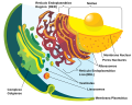
 Blausen 0350 EndoplasmicReticulum ar.png 1.440 × 1.054; 1,21 MB
-
 Blausen 0350 EndoplasmicReticulum.png 1.600 × 1.054; 1,24 MB
-
 Cellorganeller pic swe 27-05-2007.png 603 × 481; 100 KB
-
 Clara cell lung - TEM.jpg 640 × 480; 98 KB
-
 Drawing of Endoplasmic Reticulum.jpg 2.435 × 1.807; 1,19 MB
-
 Endomembrane system diagram cs.svg 638 × 530; 326 KB
-
 Endomembrane system diagram de.svg 612 × 486; 96 KB
-
 Endomembrane system diagram en.svg 612 × 486; 109 KB
-
 Endomembrane system diagram es.svg 662 × 506; 95 KB
-
 Endomembrane system diagram fr.svg 612 × 486; 244 KB
-
 Endomembrane system diagram hu.svg 638 × 530; 326 KB
-
 Endomembrane system diagram id.svg 640 × 500; 188 KB
-
 Endomembrane system diagram it.svg 612 × 486; 79 KB
-
 Endomembrane system diagram ka.svg 612 × 486; 89 KB
-
 Endomembrane system diagram ku .svg 612 × 486; 88 KB
-
 Endomembrane system diagram ln.svg 640 × 500; 188 KB
-
 Endomembrane system diagram nl.svg 765 × 600; 192 KB
-
 Endomembrane system diagram notext.svg 560 × 460; 98 KB
-
 Endomembrane system diagram pl.svg 612 × 486; 249 KB
-
 Endomembrane system diagram pt.svg 612 × 486; 286 KB
-
 Endomembrane system diagram ru.svg 612 × 486; 78 KB
-
 Endomembrane system diagram tr.svg 612 × 486; 163 KB
-
 Endomembrane system diagram uk.svg 614 × 498; 295 KB
-
 Endomembrane system diagram zh-tw.svg 612 × 486; 80 KB
-
 Endomembrane system diagram zh.svg 612 × 486; 87 KB
-
 Endoplasmic reticulum - ribosomes.jpg 1.221 × 788; 356 KB
-
 Endoplasmic reticulum -- Smart-Servier.jpg 10.240 × 5.760; 2,51 MB
-
 Endoplasmic reticulum 1 -- Smart-Servier.png 1.755 × 835; 284 KB
-
 Endoplasmic reticulum 2 -- Smart-Servier.png 1.898 × 723; 329 KB
-
 Endoplasmic reticulum 3 -- Smart-Servier.png 1.900 × 666; 282 KB
-
 Endoplasmic reticulum 4 -- Smart-Servier.png 1.760 × 843; 334 KB
-
 Endoplasmic reticulum 5 -- Smart-Servier.png 1.749 × 459; 126 KB
-
 Endoplasmic reticulum 6 -- Smart-Servier.png 2.117 × 674; 223 KB
-
 Endoplasmic reticulum 7 -- Smart-Servier.png 2.118 × 611; 170 KB
-
 Endoplasmic reticulum 8 -- Smart-Servier.png 1.753 × 490; 161 KB
-
-
-
ER-network-dynamics-are-differentially-controlled-by-myosins-XI-K-XI-C-XI-E-XI-I-XI-1-and-XI-2-Movie1.ogv 4,3 s, 1.280 × 720; 2,11 MB
-
ER-network-dynamics-are-differentially-controlled-by-myosins-XI-K-XI-C-XI-E-XI-I-XI-1-and-XI-2-Movie2.ogv 4,3 s, 1.280 × 720; 2,5 MB
-
ER-network-dynamics-are-differentially-controlled-by-myosins-XI-K-XI-C-XI-E-XI-I-XI-1-and-XI-2-Movie3.ogv 4,3 s, 1.280 × 720; 1,88 MB
-
ER-network-dynamics-are-differentially-controlled-by-myosins-XI-K-XI-C-XI-E-XI-I-XI-1-and-XI-2-Movie4.ogv 4,5 s, 1.280 × 720; 2,25 MB
-
ER-network-dynamics-are-differentially-controlled-by-myosins-XI-K-XI-C-XI-E-XI-I-XI-1-and-XI-2-Movie5.ogv 4,3 s, 1.280 × 720; 1,33 MB
-
ER-network-dynamics-are-differentially-controlled-by-myosins-XI-K-XI-C-XI-E-XI-I-XI-1-and-XI-2-Movie6.ogv 4,3 s, 1.280 × 720; 1,38 MB
-
ER-network-dynamics-are-differentially-controlled-by-myosins-XI-K-XI-C-XI-E-XI-I-XI-1-and-XI-2-Movie7.ogv 4,3 s, 1.280 × 720; 1,45 MB
-
ER-network-dynamics-are-differentially-controlled-by-myosins-XI-K-XI-C-XI-E-XI-I-XI-1-and-XI-2-Movie8.ogv 4,3 s, 1.280 × 720; 1,25 MB
-
ER-network-dynamics-are-differentially-controlled-by-myosins-XI-K-XI-C-XI-E-XI-I-XI-1-and-XI-2-Movie9.ogv 3,3 s, 1.280 × 720; 683 KB
-
 Esquema de la teoria de l'endosimbio siseriada.png 4.408 × 6.476; 2,96 MB
-
Exogenous-Ether-Lipids-Predominantly-Target-Mitochondria-pone.0031342.s006.ogv 28 s, 480 × 360; 8,69 MB
-
-
-
-
-
-
-
-
-
-
-
-
-
Induction-of-protein-body-formation-in-plant-leaves-by-elastin-like-polypeptide-fusions-1741-7007-7-48-S1.ogv 5,0 s, 1.436 × 1.099; 780 KB
-
Induction-of-protein-body-formation-in-plant-leaves-by-elastin-like-polypeptide-fusions-1741-7007-7-48-S10.ogv 15 s, 1.106 × 1.099; 3,34 MB
-
Induction-of-protein-body-formation-in-plant-leaves-by-elastin-like-polypeptide-fusions-1741-7007-7-48-S11.ogv 3,5 s, 1.106 × 1.099; 434 KB
-
Induction-of-protein-body-formation-in-plant-leaves-by-elastin-like-polypeptide-fusions-1741-7007-7-48-S2.ogv 7,3 s, 1.013 × 1.099; 948 KB
-
Induction-of-protein-body-formation-in-plant-leaves-by-elastin-like-polypeptide-fusions-1741-7007-7-48-S3.ogv 4,7 s, 1.253 × 1.099; 792 KB
-
Induction-of-protein-body-formation-in-plant-leaves-by-elastin-like-polypeptide-fusions-1741-7007-7-48-S4.ogv 6,0 s, 1.106 × 1.099; 2,77 MB
-
-
Induction-of-protein-body-formation-in-plant-leaves-by-elastin-like-polypeptide-fusions-1741-7007-7-48-S6.ogv 13 s, 1.106 × 1.099; 2,62 MB
-
Induction-of-protein-body-formation-in-plant-leaves-by-elastin-like-polypeptide-fusions-1741-7007-7-48-S7.ogv 5,0 s, 1.106 × 1.099; 3,55 MB
-
Induction-of-protein-body-formation-in-plant-leaves-by-elastin-like-polypeptide-fusions-1741-7007-7-48-S8.ogv 5,0 s, 1.106 × 1.099; 1,36 MB
-
Induction-of-protein-body-formation-in-plant-leaves-by-elastin-like-polypeptide-fusions-1741-7007-7-48-S9.ogv 11 s, 1.106 × 1.099; 4,15 MB
-
Inhibition-of-the-Unfolded-Protein-Response-Mechanism-Prevents-Cardiac-Fibrosis-pone.0159682.s006.ogv 1 min 14 s, 640 × 368; 39,48 MB
-
Inside-out-Ca2+-signalling-prompted-by-STIM1-conformational-switch-ncomms8826-s2.ogv 5,3 s, 1.160 × 448; 559 KB
-
Inside-out-Ca2+-signalling-prompted-by-STIM1-conformational-switch-ncomms8826-s3.ogv 5,0 s, 752 × 564; 208 KB
-
Inside-out-Ca2+-signalling-prompted-by-STIM1-conformational-switch-ncomms8826-s4.ogv 5,0 s, 1.004 × 504; 288 KB
-
Inside-out-Ca2+-signalling-prompted-by-STIM1-conformational-switch-ncomms8826-s5.ogv 6,0 s, 1.176 × 592; 89 KB
-
-
-
-
-
-
-

-
-
Mitochondrial-pleomorphy-in-plant-cells-is-driven-by-contiguous-ER-dynamics-Video1.ogv 20 s, 290 × 184; 1,39 MB
-
Mitochondrial-pleomorphy-in-plant-cells-is-driven-by-contiguous-ER-dynamics-Video2.ogv 3,8 s, 548 × 192; 618 KB
-
Mitochondrial-pleomorphy-in-plant-cells-is-driven-by-contiguous-ER-dynamics-Video3.ogv 7,3 s, 368 × 258; 970 KB
-
Mitochondrial-pleomorphy-in-plant-cells-is-driven-by-contiguous-ER-dynamics-Video4.ogv 21 s, 392 × 602; 1,98 MB
-
Mitochondrial-pleomorphy-in-plant-cells-is-driven-by-contiguous-ER-dynamics-Video5.ogv 48 s, 386 × 246; 4,3 MB
-
Mitochondrial-pleomorphy-in-plant-cells-is-driven-by-contiguous-ER-dynamics-Video6.ogv 45 s, 378 × 146; 3,25 MB
-
Molecular-Determinants-and-Dynamics-of-Hepatitis-C-Virus-Secretion-ppat.1002466.s016.ogv 6,2 s, 270 × 235; 145 KB
-
Molecular-Determinants-and-Dynamics-of-Hepatitis-C-Virus-Secretion-ppat.1002466.s017.ogv 10 s, 476 × 347; 584 KB
-
Molecular-Determinants-and-Dynamics-of-Hepatitis-C-Virus-Secretion-ppat.1002466.s018.ogv 3,1 s, 291 × 230; 141 KB
-
Molecular-Determinants-and-Dynamics-of-Hepatitis-C-Virus-Secretion-ppat.1002466.s019.ogv 6,7 s, 216 × 234; 391 KB
-
Molecular-Determinants-and-Dynamics-of-Hepatitis-C-Virus-Secretion-ppat.1002466.s020.ogv 5,0 s, 82 × 118; 83 KB
-
Molecular-Determinants-and-Dynamics-of-Hepatitis-C-Virus-Secretion-ppat.1002466.s021.ogv 9,6 s, 240 × 283; 166 KB
-
-
Molecular-Determinants-and-Dynamics-of-Hepatitis-C-Virus-Secretion-ppat.1002466.s023.ogv 4,1 s, 65 × 89; 26 KB
-
Molecular-Determinants-and-Dynamics-of-Hepatitis-C-Virus-Secretion-ppat.1002466.s024.ogv 9,3 s, 58 × 58; 42 KB
-
-
Molecular-Determinants-and-Dynamics-of-Hepatitis-C-Virus-Secretion-ppat.1002466.s026.ogv 4,0 s, 68 × 74; 21 KB
-
Molecular-Determinants-and-Dynamics-of-Hepatitis-C-Virus-Secretion-ppat.1002466.s027.ogv 2,5 s, 11 × 52; 10 KB
-
-
-
Molecular-Determinants-and-Dynamics-of-Hepatitis-C-Virus-Secretion-ppat.1002466.s030.ogv 5,4 s, 38 × 50; 19 KB
-
Motion-and-Flexibility-in-Human-Cytochrome-P450-Aromatase-pone.0032565.s011.ogv 56 s, 480 × 360; 3,85 MB
-
Motion-and-Flexibility-in-Human-Cytochrome-P450-Aromatase-pone.0032565.s012.ogv 1 min 5 s, 480 × 360; 4,04 MB
-
Motion-and-Flexibility-in-Human-Cytochrome-P450-Aromatase-pone.0032565.s013.ogv 52 s, 480 × 360; 6,07 MB
-
Motion-and-Flexibility-in-Human-Cytochrome-P450-Aromatase-pone.0032565.s014.ogv 1 min 5 s, 640 × 360; 15,66 MB
-
Motion-and-Flexibility-in-Human-Cytochrome-P450-Aromatase-pone.0032565.s015.ogv 1 min 36 s, 480 × 360; 8,03 MB
-
Motion-and-Flexibility-in-Human-Cytochrome-P450-Aromatase-pone.0032565.s016.ogv 1 min 38 s, 480 × 360; 6,03 MB
-
Motion-and-Flexibility-in-Human-Cytochrome-P450-Aromatase-pone.0032565.s017.ogv 2 min 51 s, 480 × 360; 14,38 MB
-
 N-linked protein glycosylation in the ER.svg 2.662 × 1.018; 164 KB
-
Nuclear-envelope-associated-endosomes-deliver-surface-proteins-to-the-nucleus-ncomms9218-s2.ogv 26 s, 768 × 576; 4,38 MB
-
Nuclear-envelope-associated-endosomes-deliver-surface-proteins-to-the-nucleus-ncomms9218-s3.ogv 36 s, 768 × 576; 2,56 MB
-
Nuclear-envelope-associated-endosomes-deliver-surface-proteins-to-the-nucleus-ncomms9218-s4.ogv 19 s, 640 × 480; 6,58 MB
-
Nuclear-envelope-associated-endosomes-deliver-surface-proteins-to-the-nucleus-ncomms9218-s5.ogv 13 s, 768 × 576; 4,99 MB
-
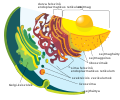
 Nucleus ER golgi ex.jpg 534 × 426; 49 KB
-
 Nucleus ER golgi.svg 492 × 565; 168 KB
-
 Nucleus ER.png 416 × 469; 31 KB
-
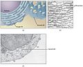
 Pancreatic acinar cells - TEM.jpg 640 × 480; 160 KB
-
 Pathologies liées aux modifications du réticulum sarcoplasmique.jpg 704 × 449; 120 KB
-
 Phosphatidylinositol hydrolysis and synthesis.jpg 6.000 × 4.200; 2,65 MB
-
 PLC.png 621 × 706; 276 KB
-
 ProteinTranscription+Synthesis.svg 957 × 531; 82 KB
-
-
-
-
-
-
-
-
-
-
-
-
-
-
-
-
 RER Gland MO.png 1.019 × 766; 1,02 MB
-
 Reticle-endoplasmatic.jpg 499 × 291; 53 KB
-
 Reticulo 3D Coanofl.png 786 × 408; 360 KB
-
 Reticulo dinámica Levadu.png 1.340 × 1.748; 1,03 MB
-
 Reticulo Endoplasmico. Inmunodetección RFP-RPGR..png 856 × 257; 359 KB
-
 Reticulo Levadura.png 357 × 432; 189 KB
-
 Reticulo Nanodominio.png 1.200 × 343; 273 KB
-
 Reticulo periferico.png 514 × 754; 378 KB
-
 Reticulo sectores Levad.png 835 × 1.341; 951 KB
-
 Retikulum stanica.png 500 × 540; 97 KB
-
 Retículo central Levadura.png 358 × 435; 229 KB
-
 RETÍCULO ENDOPLASMÁTICO.jpg 180 × 239; 37 KB
-
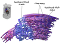
 Retîkûlûma Endoplazmî.png 762 × 418; 221 KB
-
 Rough endoplasmic reticulum.JPG 471 × 278; 33 KB
-
 Rough ER Close up.png 1.188 × 672; 138 KB
-
-
-
-
-
-
 Smooth Endoplasmic Reticulum.jpg 1.195 × 549; 194 KB
-
Spatial-Temporal-Study-of-Rab1b-Dynamics-and-Function-at-the-ER-Golgi-Interface-pone.0160838.s002.ogv 37 s, 1.920 × 1.080; 7,13 MB
-
-
Spatial-Temporal-Study-of-Rab1b-Dynamics-and-Function-at-the-ER-Golgi-Interface-pone.0160838.s004.ogv 50 s, 1.280 × 720; 4,59 MB
-
-
The-Effect-of-Gap-Junctional-Coupling-on-the-Spatiotemporal-Patterns-of-Ca2+-Signals-and-the-pcbi.1005295.s001.ogv 1 min 7 s, 250 × 103; 1,63 MB
-
The-Effect-of-Gap-Junctional-Coupling-on-the-Spatiotemporal-Patterns-of-Ca2+-Signals-and-the-pcbi.1005295.s002.ogv 1 min 7 s, 100 × 100; 2,42 MB
-
-
-
-
-
-
-
The-Effect-of-Gap-Junctional-Coupling-on-the-Spatiotemporal-Patterns-of-Ca2+-Signals-and-the-pcbi.1005295.s009.ogv 1 min 7 s, 100 × 100; 3,37 MB
-
The-Effect-of-Gap-Junctional-Coupling-on-the-Spatiotemporal-Patterns-of-Ca2+-Signals-and-the-pcbi.1005295.s010.ogv 1 min 7 s, 100 × 100; 1,56 MB
-
-
-
-
-
-
-
-
The-Human-Polyoma-JC-Virus-Agnoprotein-Acts-as-a-Viroporin-ppat.1000801.s008.ogv 9,1 s, 180 × 110; 99 KB
-
-
-
-
-
The-Intracellular-Transport-and-Secretion-of-Calumenin-12-in-Living-Cells-pone.0035344.s005.ogv 5,1 s, 512 × 512; 5,14 MB
-
The-Intracellular-Transport-and-Secretion-of-Calumenin-12-in-Living-Cells-pone.0035344.s006.ogv 5,1 s, 512 × 512; 10,09 MB
-
The-Intracellular-Transport-and-Secretion-of-Calumenin-12-in-Living-Cells-pone.0035344.s007.ogv 5,0 s, 512 × 512; 6,93 MB
-
The-Intracellular-Transport-and-Secretion-of-Calumenin-12-in-Living-Cells-pone.0035344.s008.ogv 5,0 s, 512 × 512; 13,77 MB
-
The-Intracellular-Transport-and-Secretion-of-Calumenin-12-in-Living-Cells-pone.0035344.s009.ogv 5,0 s, 512 × 512; 5,2 MB
-
The-Intracellular-Transport-and-Secretion-of-Calumenin-12-in-Living-Cells-pone.0035344.s010.ogv 5,0 s, 512 × 512; 5,49 MB


