Category:Fish parasites
Zur Navigation springen
Zur Suche springen

bei Fischen auftretende Parasiten | |||||
| Medium hochladen | |||||
| |||||
Unterkategorien
Es werden 15 von insgesamt 15 Unterkategorien in dieser Kategorie angezeigt:
In Klammern die Anzahl der enthaltenen Kategorien (K), Seiten (S), Dateien (D)
A
C
- Cymothoa exigua (9 D)
- Cystobranchus respirans (2 D)
D
E
H
- Hamacreadium cribbi (5 D)
I
L
- Lepeophtheirus salmonis (6 D)
- Lernaeenicus sprattae (1 D)
M
- Myxobolus cerebralis (16 D)
P
- Phrixocephalus cincinnatus (1 D)
T
Medien in der Kategorie „Fish parasites“
Folgende 200 Dateien sind in dieser Kategorie, von 368 insgesamt.
(vorherige Seite) (nächste Seite)-
 003 Anisakis nematode parasites in mackerel fish caught in Norway.jpg 3.030 × 2.020; 2 MB
-

-

-
 A. ocellatum infected gills in European sea bass.png 453 × 348; 454 KB
-

-

-

-
-
 Anilocra capensis.jpg 1.024 × 766; 318 KB
-

-
 Ascarophis richeri (Nematoda, Cystidicolidae).png 3.085 × 4.369; 3,81 MB
-
 Betty in mouth.jpg 599 × 762; 30 KB
-
 Bolbophorus sp on ictalurus punctatus.jpg 3.072 × 2.028; 3,06 MB
-
 California fish and game (19890788064).jpg 2.095 × 3.328; 577 KB
-
 Chimaericola leptogaster (Monogenea).png 1.579 × 2.337; 88 KB
-
 Cichlidogyrus evikae (Monogenea, Ancyrocephalidae).gif 567 × 302; 77 KB
-
 Cichlidogyrus justinei (Monogenea, Ancyrocephalidae).gif 567 × 318; 80 KB
-
 Copépode parasite (Penella filosa à confirmer).JPG 1.600 × 1.090; 1,41 MB
-
 Cryptocotyle 1.jpg 1.896 × 1.464; 149 KB
-
 Cryptocotyle 2.jpg 2.240 × 1.472; 187 KB
-
 Cryptocotyle 3.jpg 2.272 × 1.776; 167 KB
-
 CystobranchusRespiransRutilusRutilus.JPG 2.560 × 1.920; 1,82 MB
-
 Dorsal view of the head of the adult female of Ceratothoa oestroides. 02.jpg 2.272 × 1.704; 895 KB
-
 Expn0031 (14318884118).jpg 1.920 × 1.080; 1,22 MB
-
 Expn0033 (14525596393).jpg 1.920 × 1.080; 1,03 MB
-
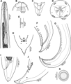
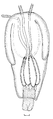
 FIG11 Nybelinia basimegacantha Tentacle.png 1.000 × 2.252; 197 KB
-
 FMIB 33711 Olypea tyrannus; Oniscus pragustator.jpeg 1.233 × 772; 122 KB
-

-

-
 FMIB 41566 Young Brook Trout, Diseased--Gill of Large Fish with Copepod Parasites.jpeg 1.713 × 1.325; 690 KB
-
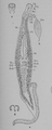
 FMIB 46116 Parasite on the Skate (Hirudo muricata).jpeg 587 × 449; 60 KB
-
 FMIB 46376 Sunfish.jpeg 1.016 × 794; 567 KB
-
 FMIB 46490 Cymothoa astrum, an Isopod parasite of fish.jpeg 532 × 691; 131 KB
-
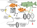
 FMIB 47807 Life cycle of the parasite.jpeg 977 × 985; 180 KB
-

-
-
 FMIB 51771 Median segments of Dib tyhrium e rdiceps.jpeg 367 × 229; 24 KB
-

-
 Gastrocotyle trachuri.jpg 1.300 × 1.030; 89 KB
-

-

-

-

-
 Goto 1894 - Studies on the Ectoparasitic Trematodes of Japan - Plate 1.png 5.424 × 3.964; 31,8 MB
-
 Goto 1894 - Studies on the Ectoparasitic Trematodes of Japan - Plate 2 Microcotyle chiri.png 1.848 × 3.067; 8,32 MB
-

-

-

-
 Goto 1894 - Studies on the Ectoparasitic Trematodes of Japan - Plate 2.png 5.412 × 4.001; 31,82 MB
-

-
 Goto 1894 - Studies on the Ectoparasitic Trematodes of Japan - Plate 3.png 4.093 × 5.696; 34,17 MB
-
 Goto 1894 - Studies on the Ectoparasitic Trematodes of Japan - Plate 4.png 3.857 × 5.637; 39,11 MB
-
 Henneguya salminicola in flesh of coho salmon, BC, Canada.JPG 1.500 × 1.125; 399 KB
-
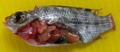
 Infected goby.jpg 734 × 332; 50 KB
-

-

-

-

-

-
-

-

-

-

-

-

-
-

-
 Journal.pone.0171392.g002 - Pseudorhabdosynochus riouxi from Mycteroperca marginata.png 1.625 × 2.400; 693 KB
-
-
 Journal.pone.0171392.g004 - Pseudorhabdosynochus bouaini from Mycteroperca costae.png 2.200 × 1.929; 557 KB
-

-
 Journal.pone.0171392.g006 - Pseudorhabdosynochus enitsuji from Mycteroperca costae.png 2.200 × 1.574; 646 KB
-

-

-
-

-

-
 Journal.pone.0171392.g012 - Pseudorhabdosynochus sinediscus from Mycteroperca costae.png 2.200 × 1.858; 485 KB
-

-

-
 Journal.pone.0171392.g015.png 2.256 × 1.831; 146 KB
-
 Journal.pone.0171392.g016.png 2.256 × 2.511; 226 KB
-
 Journal.pone.0171392.g017.png 2.257 × 2.331; 240 KB
-
 Journal.pone.0171392.g018.png 2.635 × 2.004; 242 KB
-
 Journal.pone.0171392.g019.png 2.256 × 2.106; 223 KB
-
 Journal.pone.0171392.g020.png 2.256 × 1.489; 148 KB
-
 Journal.pone.0171392.g021.png 2.256 × 1.487; 206 KB
-
 Lagenivaginopseudobenedenia sp. (Monogenea, Capsalidae).png 944 × 938; 1 MB
-
 Lamprey attached.png 784 × 349; 141 KB
-
 Lepotrema (Lepocreadiidae, Digenea) 11230 2018 9821 Fig01.png 785 × 854; 102 KB
-
 Lepotrema (Lepocreadiidae, Digenea) 11230 2018 9821 Fig02.png 785 × 831; 134 KB
-
 Lepotrema (Lepocreadiidae, Digenea) 11230 2018 9821 Fig03--07.png 785 × 1.524; 1,58 MB
-
 Lepotrema (Lepocreadiidae, Digenea) 11230 2018 9821 Fig08--09.png 785 × 1.008; 1,31 MB
-
 Lepotrema (Lepocreadiidae, Digenea) 11230 2018 9821 Fig10--14.png 785 × 1.135; 1,46 MB
-
 Lepotrema (Lepocreadiidae, Digenea) 11230 2018 9821 Fig15--21.png 785 × 1.103; 1,42 MB
-
 Lepotrema (Lepocreadiidae, Digenea) 11230 2018 9821 Fig22-28.png 785 × 1.060; 1,27 MB
-
 Lepotrema (Lepocreadiidae, Digenea) 11230 2018 9821 Fig29--35.png 785 × 1.042; 1,36 MB
-
 Lepotrema (Lepocreadiidae, Digenea) 11230 2018 9821 Fig36--39.png 785 × 1.466; 1,73 MB
-
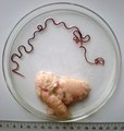
 Lernaeocera branchialis.jpg 1.024 × 768; 1,23 MB
-
 Life cycle of Amyloodinium ocellatum.png 1.184 × 642; 1,1 MB
-
 Life cycle of Huffmanela huffmani Moravec, 1987.png 1.950 × 1.479; 786 KB
-

-
 Microcotyle chrysophrii - Plate XV in Van Beneden & Hesse, 1863.png 1.324 × 1.810; 924 KB
-
 Microcotyle donavini (Microcotylidae) Atrium (Euzet & Marc).png 641 × 561; 128 KB
-
 Microcotyle donavini (Microcotylidae) Body (Euzet & Marc).png 746 × 897; 219 KB
-
 Microcotyle donavini (Microcotylidae) Clamps (Euzet & Marc).png 704 × 279; 35 KB
-
 Microcotyle donavini (Microcotylidae) Lappet (Euzet & Marc).png 374 × 315; 27 KB
-

-
 Microcotyle donavini (Microcotylidae) Oncomiracidium (Euzet & Marc).png 356 × 702; 117 KB
-
 Microcotyle isyebi body (Bouguerche, Gey, Justine & Tazerouti).jpg 121 × 446; 39 KB
-

-
-
-
 Moravec & Justine - Euterranova n. gen. and Neoterranova n. gen - parasite200141-fig2.png 3.484 × 4.179; 4,98 MB
-
 Moravec & Justine - Euterranova n. gen. and Neoterranova n. gen - parasite200141-fig3.png 3.484 × 4.179; 6,26 MB
-

-
 Moravec & Justine - Euterranova n. gen. and Neoterranova n. gen - parasite200141-fig5.png 3.484 × 2.788; 4,64 MB
-
 Moravec & Justine - Euterranova n. gen. and Neoterranova n. gen - parasite200141-fig6.png 3.484 × 3.136; 4,64 MB
-
 Moravec & Justine - New Cucullanidae - parasite200044-fig01.png 7.087 × 9.226; 4,05 MB
-
 Moravec & Justine - New Cucullanidae - parasite200044-fig02.png 3.445 × 4.132; 5,22 MB
-
 Moravec & Justine - New Cucullanidae - parasite200044-fig03.png 3.504 × 2.804; 3,32 MB
-
 Moravec & Justine - New Cucullanidae - parasite200044-fig04.png 2.959 × 3.457; 770 KB
-
 Moravec & Justine - New Cucullanidae - parasite200044-fig05.png 3.189 × 4.142; 5,17 MB
-
 Moravec & Justine - New Cucullanidae - parasite200044-fig06.png 1.581 × 2.510; 1,09 MB
-
 Moravec & Justine - New Cucullanidae - parasite200044-fig07.png 2.959 × 2.500; 561 KB
-
 Moravec & Justine - New Cucullanidae - parasite200044-fig08.png 3.445 × 4.133; 4,63 MB
-
 Moravec & Justine - New Cucullanidae - parasite200044-fig09.png 2.959 × 2.862; 667 KB
-
 Moravec & Justine - New Cucullanidae - parasite200044-fig10.png 3.445 × 4.133; 4,9 MB
-
 Moravec & Justine - New Cucullanidae - parasite200044-fig11.png 3.504 × 2.806; 3,42 MB
-
 Moravec & Justine - New Cucullanidae - parasite200044-fig12.png 2.959 × 2.970; 721 KB
-
 Moravec & Justine - New Cucullanidae - parasite200044-fig13.png 3.189 × 4.142; 4,53 MB
-
 Moravec & Justine - New Cucullanidae - parasite200044-fig14.png 3.504 × 2.806; 3,82 MB
-
 Moravec & Justine - New Cucullanidae - parasite200044-fig15.png 1.581 × 1.808; 397 KB
-
 Moravec & Justine - New Cucullanidae - parasite200044-fig16.png 1.280 × 1.538; 339 KB
-
 Moravec & Justine - New Cucullanidae - parasite200044-fig17.png 1.280 × 1.745; 321 KB
-
 Moravec & Justine Anisakidae 2020 parasite200028-fig01.png 3.012 × 2.196; 463 KB
-
 Moravec & Justine Anisakidae 2020 parasite200028-fig02.png 2.835 × 3.402; 4,35 MB
-
 Moravec & Justine Anisakidae 2020 parasite200028-fig03.png 3.012 × 3.454; 628 KB
-
 Moravec & Justine Anisakidae 2020 parasite200028-fig04.png 2.835 × 3.402; 4,56 MB
-
 Moravec & Justine Anisakidae 2020 parasite200028-fig05.png 2.835 × 2.268; 2,3 MB
-
 Moravec & Justine Anisakidae 2020 parasite200028-fig06.png 3.012 × 2.441; 473 KB
-
 Moravec & Justine Anisakidae 2020 parasite200028-fig07.png 2.835 × 3.402; 3,9 MB
-
 Moravec & Justine Anisakidae 2020 parasite200028-fig08.png 1.417 × 2.268; 1,36 MB
-
 Moravec & Justine Anisakidae 2020 parasite200028-fig09.png 3.012 × 3.169; 638 KB
-
 Moravec & Justine Anisakidae 2020 parasite200028-fig10.png 2.835 × 3.402; 3,94 MB
-
 Moravec & Justine Anisakidae 2020 parasite200028-fig11.png 2.835 × 3.685; 4,41 MB
-
 Moravec & Justine Anisakidae 2020 parasite200028-fig12.png 3.012 × 3.477; 843 KB
-
 Moravec & Justine Anisakidae 2020 parasite200028-fig13.png 3.154 × 3.784; 4,57 MB
-
 Moravec & Justine Anisakidae 2020 parasite200028-fig14.png 2.835 × 2.268; 2,43 MB
-
 Moravec & Justine Anisakidae 2020 parasite200028-fig15.png 3.012 × 3.307; 711 KB
-
 Moravec & Justine Anisakidae 2020 parasite200028-fig16.png 2.835 × 3.685; 4,67 MB
-
 Moravec & Justine Anisakidae 2020 parasite200028-fig17.png 2.835 × 2.268; 2,35 MB
-
 Moravec & Justine Spirurida 2020 parasite190153-fig1.png 2.662 × 3.137; 1,28 MB
-
 Moravec & Justine Spirurida 2020 parasite190153-fig2.png 2.662 × 2.131; 2,76 MB
-
 Moravec & Justine Spirurida 2020 parasite190153-fig3.png 1.422 × 2.542; 546 KB
-
 Moravec & Justine Spirurida 2020 parasite190153-fig4.png 2.662 × 3.672; 1,45 MB
-
 Moravec & Justine Spirurida 2020 parasite190153-fig5.png 2.662 × 3.194; 3,84 MB
-
 Moravec & Justine Spirurida 2020 parasite190153-fig6.png 2.662 × 3.459; 3,47 MB
-
 Moravec & Justine Spirurida 2020 parasite190153-fig7.png 2.662 × 3.164; 955 KB
-
 Moravec & Justine Spirurida 2020 parasite190153-fig8.png 2.657 × 3.188; 3 MB
-
 Moravec & Justine Spirurida 2020 parasite190153-fig9.png 2.662 × 2.131; 2,18 MB
-
 NIE 1905 Menhaden - fish-louse of the menhaden.jpg 455 × 92; 8 KB
-
 Parasite 20,42(2013) Complete life cycle of a pennellid Peniculus minuticaudae -fig1.tif 2.067 × 2.933; 686 KB
-
 Parasite 20,42(2013) Complete life cycle of a pennellid Peniculus minuticaudae -fig2.tif 1.654 × 1.141; 753 KB
-
 Parasite 20,42(2013) Complete life cycle of a pennellid Peniculus minuticaudae -fig3.tif 2.067 × 2.902; 572 KB
-
 Parasite 20,42(2013) Complete life cycle of a pennellid Peniculus minuticaudae -fig4.tif 2.067 × 2.885; 590 KB
-
 Parasite 20,42(2013) Complete life cycle of a pennellid Peniculus minuticaudae -fig5.tif 2.067 × 2.901; 631 KB
-
 Parasite 20,42(2013) Complete life cycle of a pennellid Peniculus minuticaudae -fig6.tif 2.067 × 2.920; 687 KB
-
 Parasite 20,42(2013) Complete life cycle of a pennellid Peniculus minuticaudae -fig7.tif 2.157 × 3.100; 1.004 KB
-
 Parasite 20,42(2013) Complete life cycle of a pennellid Peniculus minuticaudae -fig8.tif 2.343 × 1.950; 613 KB
-

-
-

-

-
-

-

-
-

-
 Parasite140007-fig9 Philometra selaris Moravec & Justine, 2014 (Nematoda, Philometridae).tif 2.343 × 2.811; 4,14 MB
-
 Parasite140042-fig2 Clinostomum phalacrocoracis metacercariae in fish.tif 1.654 × 722; 1,49 MB
-

-
 Parasite140092-fig2 FIG 3 Cestoda Trypanorhyncha melanized plerocerci in Epinephelus sp.JPG 3.264 × 2.448; 2,44 MB
-

-
 Parasite140092-fig2 FIGS2-7 Metacestodes of trypanorhynch cestodes Photos.png 2.008 × 2.128; 4,65 MB
-
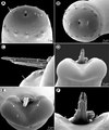
 Parasite140092-fig3 - FIG10 Nybelinia sp C.png 900 × 1.656; 186 KB
-
 Parasite140092-fig3 - FIG11 Nybelinia basimegacantha body.png 908 × 1.744; 161 KB
-
 Parasite140092-fig3 - FIG8 Nybelinia sp A.png 845 × 1.679; 177 KB
-
 Parasite140092-fig3 - FIG9 Nybelinia sp B.png 737 × 1.648; 161 KB
-
 Parasite140092-fig3 T FIGS 8-11 Metacestodes of trypanorhynch cestodes Drawings.png 3.832 × 4.389; 1,49 MB
-
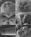
 Parasite140121-fig1 Pseudorhabdosynochus jeanloui (Monogenea, Diplectanidae) Fig1.png 2.200 × 2.184; 781 KB
-
-
-

-

-

-
 Parasite140121-fig2 Pseudorhabdosynochus jeanloui (Monogenea, Diplectanidae) Fig2.png 2.176 × 2.180; 758 KB
-

-


_Body_(Bouguerche,_Gey,_Tazerouti_%26_Justine).png)
_Clamps_(Bouguerche,_Gey,_Tazerouti_%26_Justine).png)
_Genital_atrium_(Bouguerche,_Gey,_Tazerouti_%26_Justine).png)
_Terminal_lappet_(Bouguerche,_Gey,_Tazerouti_%26_Justine).png)

_(14568749128).jpg)
.png)


.jpg)
.png)
.gif)
.gif)
.JPG)


.jpg)
.jpg)


.jpeg)


_Specimen_from_Tasman_Sea,_having_parasitic_lernaean_crustaceans,_to.jpeg)


.png)

_11230_2018_9821_Fig01.png)
_11230_2018_9821_Fig02.png)
_11230_2018_9821_Fig03--07.png)
_11230_2018_9821_Fig08--09.png)
_11230_2018_9821_Fig10--14.png)
_11230_2018_9821_Fig15--21.png)
_11230_2018_9821_Fig22-28.png)
_11230_2018_9821_Fig29--35.png)
_11230_2018_9821_Fig36--39.png)


_in_MacCallum_1915_Notes_on_the_genus_Microcotyle.png)

_Atrium_(Euzet_%26_Marc).png)
_Body_(Euzet_%26_Marc).png)
_Lappet_(Euzet_%26_Marc).png)
_Oncomiracidium_%26_Egg_(Euzet_%26_Marc).png)
_Oncomiracidium_(Euzet_%26_Marc).png)
_in_MacCallum_1915_Notes_on_the_genus_Microcotyle.png)


_Fig1.png)
_Fig1b_MCO.png)
_Fig1b%26c_Sclerotized_organs.png)
_Fig1c_Vagina.png)
_Fig1d_Reroductive_organs.png)
_Fig2.png)
_Clamps_(Euzet_%26_Marc).png)
.jpg)
_in_MacCallum_1915_Notes_on_the_genus_Microcotyle.png)
_Fig1a_body.png)
_Fig2a%26b_Squamodics.png)
_Fig2c-i_Sclerotised_parts_of_haptor.png)