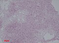
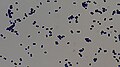
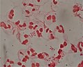
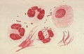
Category:Gram stains
नेविगेशन पर जाएँ
खोज पर जाएँ
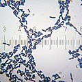
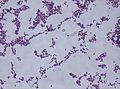
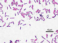
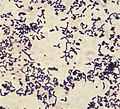
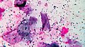
English: Gram staining (or Gram's method) is an empirical method of differentiating bacterial species into two large groups (Gram-positive and Gram-negative) based on the chemical and physical properties of their cell walls.
The method is named after its inventor, the Danish scientist Hans Christian Gram (1853 – 1938), who developed the technique in 1884 to discriminate between pneumococci and Klebsiella pneumoniae bacteria.
Deutsch: Gram-Färbung ist eine empirische Methode zur Unterscheidung von Bakterienarten in zwei große Gruppen (Gram-positive und Gram-negative) auf der Grundlage der chemischen und physikalischen Eigenschaften ihrer Zellwände. Durch das Färbeverfahren erscheinen im Lichtmikroskop grampositive Bakterien blau, gramnegative rot. Die Methode ist nach ihrem Erfinder, dem dänischen Wissenschaftler Hans Christian Gram (1853 - 1938) benannt, der 1884 die Technik entwickelte, um zwischen Pneumokokken und Klebsiella pneumoniae Bakterien zu unterscheiden.
microbiological method for identification; method of staining used to differentiate bacterial species into two large groups (gram-positive and gram-negative)  | |||||
| मीडिया अपलोड करें | |||||
| जिसका उदाहरण है |
| ||||
|---|---|---|---|---|---|
| जिसका उपवर्ग है | |||||
| द्वारा नामांकित | |||||
| खोजकर्ता या आविष्कारक | |||||
| |||||
उपश्रेणियाँ
इस श्रेणी की कुल ४ में से ४ उपश्रेणियाँ निम्नलिखित हैं।
G
S
"Gram stains" श्रेणी में मीडिया
इस श्रेणी की कुल १३५ में से १३५ चित्र निम्नलिखित हैं।
-

-

-
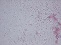
 11G0002 lores.jpg ७०० × ४७०; ७८ KB
-
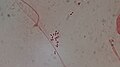
 20100905 205851 Streptobacilli.jpg १,६०० × ९८९; १२९ KB
-
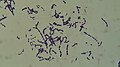
 20100905 211652 SpirochetesZoom.jpg १,५७७ × १,१४६; ३६९ KB
-
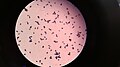
 20101017 175758 Bacilli.jpg ३,०७२ × २,३०४; २९० KB
-
 20101017 175839 BacilliFromSqwincher.jpg १,६०० × १,६००; १९३ KB
-
 20101017 231210 Staphylococcus.jpg १,५०० × १,५००; २२१ KB
-
 20101210 020132 StreptococcusThermophilus.jpg १,४०० × १,०००; ७८ KB
-
 20101212 205549 LactobacillusAcidophilus.jpg ३,०७२ × २,३०४; ३२७ KB
-
 20110322 193829 Bacteria SMB UTI.jpg १,६०० × १,६००; २५८ KB
-
 Aerococcus urinae - microscopy.jpg १,९२० × १,०८०; ७०७ KB
-
 Aeromonas veronii biovar sobria Gram Stain on Microscope Slide.jpg १,७९६ × १,७९६; ३६४ KB
-
 Bacillus anthracis Gram.jpg २,८९२ × १,९४०; ६.४६ MB
-
 Bacillus cereus Gram.jpg ६०० × ४५०; १७ KB
-
 Bacillus coagulans 01.jpg ७०० × ४६०; ५६ KB
-
 Bacillus subtilis gram stain CDC PHIL 19261.jpg ७०० × ४६०; १९ KB
-
 Bacillus subtilis Gram stain.jpg २,०८० × १,५३६; २.३१ MB
-
 Bacillus subtilis Gram.jpg ५०० × ३७५; ३८ KB
-
 Bacterial cell wall.png ६०० × ४०५; १०५ KB
-
 Bacterial vaginosis Gram stain.jpg ५६९ × ८०४; ८२८ KB
-
 BacteroidesFragilis Gram.jpg ७०० × ४६८; ४८ KB
-
 Beta-hemoltic streptococci in Gram stain.jpg ४,००० × ३,०००; १.०३ MB
-
 Bifidobacterium adolescentis Gram.jpg ६०० × ४५०; ३५ KB
-
 Bordetella pertussis.jpg २,५८८ × २,३८३; २.१५ MB
-
 Brucella melitensis.jpg २,८८० × २,१८५; १.०६ MB
-
 Brucella spp.JPG २,८३५ × २,२५७; २.४२ MB
-
 C tetani gram stain.jpg ४४० × २९८; २१ KB
-
 Candida dubliniensis.jpg १,६०० × १,२००; ५८६ KB
-
 Citrobacter freundii Gram stain.jpg २,०८० × १,५३६; २.२ MB
-
 Clostridi bacteria gram coloration.jpg २,४२५ × २,४२४; १.६३ MB
-
 Clostridium perfringens gas gangrene.jpg १,२०० × ८९०; १९८ KB
-
 Clostridium perfringens.jpg १,८१३ × १,२०६; २.६४ MB
-
 Corynebacterium xerosis Gram stain.jpg ७२० × ५४२; ७२ KB
-
 Diphtheroides.jpg १,९१२ × १,१०८; ९३६ KB
-
 Diphtheroids as pathogens in Gram staining of sputum.jpg ४,००० × २,२५०; १.०६ MB
-
 E choli Gram.JPG १,६०९ × १,४६१; ८७२ KB
-
 E.coli gram stain.jpg ४,०३२ × ३,०२४; १.८८ MB
-
 Encapsulated strain of Streptococcus pneumoniae in clinical sample sputum Gram staining.jpg ४,१६० × २,३४०; २.८२ MB
-
 Enterobacter aerogenes.jpg २,०८० × १,५३६; २.१९ MB
-
 Enterococcus histological pneumonia 01.png ५९७ × ४३४; ४१६ KB
-
 Escherichia coli Gram.jpg ५०० × ३७५; ३६ KB
-
 Gasbrand02.JPG १,६०० × १,२००; १८४ KB
-
 Gonococcal urethritis PHIL 4085 lores.jpg ३,६०४ × २,३७६; ३.५ MB
-
 Gram - algorithm.png १,०८९ × ५१८; ३५ KB
-
 Gram -Positive and Negative Bacteria.jpg ४,००० × २,२५०; १.१६ MB
-
 Gram escherichia coli und micrococcus luteus.jpg ३८७ × २९०; ३८ KB
-
 Gram Negative bacilli of Klebsiella in Gram stained smear of culture.jpg ४,००० × २,२५०; १.८१ MB
-
 Gram Negative Rods and Pus cells in Gram staining.jpg ४,००० × २,२५०; १,००२ KB
-
 Gram Negative Rods in Sputum Gram Staining.jpg ४,००० × २,२५०; १ MB
-
 Gram Negative Rods of Aeromonas hydrophila.jpg ४,००० × २,२५०; १.०८ MB
-
 Gram negative rods.jpg ४,००० × ३,०००; १.३२ MB
-
 Gram Positive Classification.png ६६७ × ४५८; ६९ KB
-
 Gram positive cocci in chains of Streptococcus agalactiae.jpg ४,००० × २,२५०; १.४ MB
-
 Gram positive cocci in singles, pairs and chains.jpg ४,००० × २,२५०; १.१९ MB
-
 Gram positive rods of Diphtheroid.jpg ४,००० × ३,०००; १.२७ MB
-
 Gram positive yeast cells in Gram staining of culture.jpg १,९२० × १,०८०; २८८ KB
-
 Gram Stain Anthrax.jpg ६०० × ४०५; ४१ KB
-
 Gram stain of Rothia dentocariosa.jpg १,११२ × १,००९; २५७ KB
-
 Gram stain of Streptococcus pneumoniae.jpg २,९८० × २,३२२; १.३८ MB
-
 Gram stain saliva.jpg १,१७९ × ६५७; ६७८ KB
-
 Gram Stain.png १,२८० × ७२०; २२१ KB
-
 Gram Stained Smear of Sputum for Observation.jpg ४,००० × ३,०००; ५.७८ MB
-
 Gram Staining Bacteria.jpg १,९२० × १,०८०; ७९३ KB
-
 Gram variability.jpg ९३९ × ४९२; २४९ KB
-
 Gram- negative bacteria.jpg ४५२ × ६०४; ९४ KB
-
 Gram-negative Bacteria - Lab methods algorithm.svg १,३५० × ६००; १६ KB
-
 Gram-positive stain.jpg ८०० × ५३३; ८३ KB
-
 Gram-Stained Yogurt Bacteria.jpg ९,००० × १२,०००; ६.४९ MB
-
 Gram-staining-of-D.jpg ६०० × ४४९; ९८ KB
-
 Haemophilus influenzae Gram.JPG १,९०२ × १,५९१; १.१५ MB
-
 Haemophilus influenzae sputum 1000x edited.jpg १,९२० × १,२००; ९१५ KB
-
 Ideal smear of Sputum.jpg ४,००० × ३,०००; २.६४ MB
-
 Klebsiella oxytoca.jpg २,०८० × १,५३६; २.१९ MB
-
 Lactobacilli (Gram stain).jpg ३,००८ × २,०००; २.१३ MB
-
 Lactobacillus sp 01.png ६२५ × ४९८; २५७ KB
-
 LegionellaPneumophila Gram.jpg ७०० × ४७५; ५४ KB
-
 Leuconostoc mesenteroides Gram Staining.jpg ४,००० × ३,०००; १.३२ MB
-
 LF and NLF Gram Negative Bacteria on MacConkey medium.jpg २,३४० × ४,१६०; २.८३ MB
-
 Methylobacterium (Gram stain).jpg २,०८० × १,५३६; २.०९ MB
-
 Microbiology gram stain.jpg ४,०३२ × ३,०२४; २.३१ MB
-
 Micrococcus in Gram stain.jpg ४,००० × २,२५०; १.०५ MB
-
 Neisseria gonorrhoeae and pus cells.jpg ३,६०४ × २,९२५; १.०९ MB
-
 Neisseria gonorrhoeae diplococci inside a neutrophil.jpg २,२३२ × १,६४५; ३५७ KB
-
 Neisseria gonorrhoeae PHIL 3693 lores.jpg १,७७४ × १,१५८; ५१७ KB
-
 Neisseria gonorrhoeae.jpg ५,३१२ × २,९८८; २.८५ MB
-
 Neisseria meningitidis CSF Gram 1000.jpg १,९२० × १,२००; ६३९ KB
-
 Nocardia in Gram Stain.tif १,३६० × १,०२४; ३.९९ MB
-
 Nocardiosis - Gram stain Case 149 (5286067518).jpg १,२८० × ९६०; ६४६ KB
-
 Nocardiosis - Gram stain Case 149 (5286067644).jpg १,२८० × ९६०; ६६१ KB
-
 Non-Ideal smear of Sputum.jpg ४,००० × ३,०००; २.३ MB
-
 Normal flora, Pus cells and Epithelial cells.jpg ४,००० × ३,०००; १.३९ MB
-
 Normal Upper Respiratory Tract Flora in Gram Stained smear of Sputum.jpg ४,००० × ३,०००; १.३९ MB
-
 Numerous Gram Negative Bacteria and Pus cells in Gram staining of sputum.jpg ४,००० × २,२५०; १,०२० KB
-
 Plenty of pus cells and Gram negative diplococci in Gram stained smear of sputum.jpg ४,१६० × २,३४०; १.४६ MB
-
 Pseudomonas aeruginosa Gram.jpg ५०० × ३७५; ६६ KB
-
 Pseudomonas aeruginosa gram.jpg ५९० × ४९९; ५० KB
-
 Pseudomonas aeruginosa smear Gram 2010-02-10.JPG १,३४२ × १,००६; ६४९ KB
-
 Pseudomonas fluorescens Gram Stain on Microscope Slide.jpg १,२०० × १,२००; ४५४ KB
-
 Pseudomonas fluorescens.jpg २,०८० × १,५३६; २.३५ MB
-
 Purulent inflammation, Gram stain 3.jpg १,९२० × १,२८०; १.४७ MB
-
 Pus cells with Neisseria gonorrhoeae.jpg ३,००८ × २,०००; २.१३ MB
-
 Pus cells.jpg ४,००० × ३,०००; ५६३ KB
-
-
 Rhodococcus fascians.jpg २,०८० × १,५३६; २.२१ MB
-
 Rothia dentocariosa PHIL15195.png ३,०४५ × २,००५; १३.२९ MB
-
 Rothia dentocariosa PHIL21290.png ३,०४५ × २,००५; १२.३६ MB
-
 Rothia dentocariosa PHIL21292.png ३,०४५ × २,००५; १४.६४ MB
-
 Rothia dentocariosa PHIL21293.png ३,०४५ × २,००५; १५.७ MB
-
 Rothia dentocariosa PHIL21294.png ३,०४५ × २,००५; १४.३१ MB
-
 Salmonella Typhimurium Gram.jpg ५०० × ३७५; ८४ KB
-
 Shigella flexneri Gram Stain on Microscope Slide.jpg १,२२० × १,२२०; ४६९ KB
-
 Shigella flexneri Gram.jpg ५०० × ३७५; ३० KB
-
 Spermatozoa in Gram Stained Smear of Semen.jpg ४,००० × २,२५०; १.०४ MB
-
 Staph sputum.JPG १,५८४ × १,५२३; ९२९ KB
-
 Staphylococcus aureus Gram.jpg ५०० × ३७५; २४ KB
-
 Staphylococcus saprophyticus.jpg २,०८० × १,५३६; २.३ MB
-
 Stenotrophomonas maltophilia.jpg २,०८० × १,५३६; २.१४ MB
-
 Streptobacilli and streptococci in Gram stained smear microscopy at 1000X magnification.jpg ४,१६० × २,३४०; १.२३ MB
-
 Streptococcus mutans 01.jpg ६५८ × ४८१; ८७ KB
-
 Streptococcus mutans Gram.jpg ६०० × ४५०; ४१ KB
-
 StreptococcusMutans.jpg ६५४ × ४७३; ६३ KB
-
 The Gram Staining - Bacteria Gram Negative.JPG २,४४८ × ३,२६४; १.९२ MB
-
 Vibrio cholerae gram stain CDC.jpg ७०० × ७२३; ५६ KB
-
 Viridans strptococci in Gram stain.jpg ४,००० × ३,०००; १.२४ MB
-
 Yeast cells in Gram stained smear of sputum.jpg ४,००० × २,२५०; २.२४ MB
-
 Yersinia enterocolitica gram.jpg १,४०० × ९३९; ४५९ KB
-
 Кал ребенок лактобациллы.jpg १,२८० × ९६०; १.२५ MB
-
 Нитчасті бактерії 01.jpg १,८३६ × २,४४८; १,०२३ KB
-
 Нитчасті бактерії 02.jpg १,८३६ × २,४४८; १.०८ MB
-
 Нитчасті бактерії 03.jpg १,८३६ × ३,२६४; १.११ MB
-
 Нитчасті бактерії 04.jpg १,८३६ × ३,२६४; १.२४ MB
-
 Нитчасті бактерії 05.jpg १,८३६ × ३,२६४; १.५४ MB
-
 Нитчасті бактерії 06.jpg १,८३६ × ३,२६४; १.५५ MB
-
 Плазмоциты в кале.jpg १,२८० × ९६०; १.४६ MB


.jpg)


.jpg)


.jpg)
.jpg)


