Category:Gyri
Salti al navigilo
Salti al serĉilo
la faldoj de la kortekso signife pliigas ĝian surfacareon kompare kun glata kortekso de la sama grandeco. La faldoj estis kreitaj en la evolua procezo dum kiu la volumeno de la cerba kortekso pliiĝis multe pli ol la volumeno de la krania kavo | |||||
| Alŝuti plurmedion | |||||
| Estas |
| ||||
|---|---|---|---|---|---|
| Subaro de |
| ||||
| Parto de | |||||
| |||||

see also
and
Subkategorioj
Ĉi tiu kategorio havas la 28 jenajn subkategoriojn, el 28 entute.
- SVG gyri (2 D)
A
- Angular gyrus (29 D)
C
- Cuneus (22 D)
F
G
H
- Heschl's gyrus (32 D)
I
- Inferior temporal gyrus (30 D)
L
- Lateral occipital gyrus (9 D)
M
- Middle frontal gyrus (32 D)
- Middle temporal gyrus (25 D)
O
P
- Paracentral lobule (22 D)
- Planum temporale (15 D)
- Postcentral gyrus (42 D)
- Precentral gyrus (40 D)
- Precuneus (33 D)
S
- Straight gyrus (18 D)
- Subcallosal area (5 D)
- Superior frontal gyrus (44 D)
- Superior parietal lobule (18 D)
- Supramarginal gyrus (28 D)
Dosieroj en kategorio “Gyri”
La jenaj 200 dosieroj estas en ĉi tiu kategorio, el 204 entute.
(antaŭa paĝo) (sekva paĝo)-
"Traite complet de l'anatomie...",Foville, 1844 Wellcome L0019133.jpg 1 230 × 1 557; 552 KB
-
"Traite complet de l'anatomie...",Foville, 1844 Wellcome L0019135.jpg 3 763 × 4 947; 4,58 MB
-
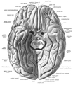
04 2 facies ventralis cerebri gyri.jpg 1 799 × 2 122; 644 KB
-
A human head dissected; "In memoriam". Wellcome L0074866.jpg 4 817 × 5 611; 4,43 MB
-
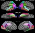
Brain parcellation.jpg 600 × 320; 81 KB
-
Brain surface by Raymond Vieussens, 1684.png 455 × 386; 220 KB
-
Brain Surface Gyri.SVG 1 024 × 731; 28 KB
-
Brain visualization.jpg 500 × 500; 40 KB
-
Brain-1.jpg 484 × 356; 58 KB
-
Brain-disease-gyrification.png 1 794 × 550; 620 KB
-
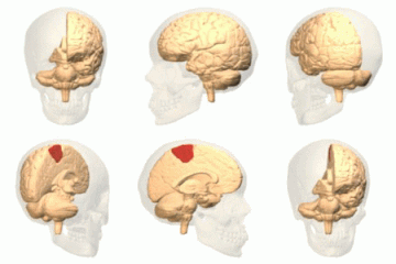
Brodmann area 3 1 2.png 300 × 190; 20 KB
-
Causes of Autism in Brain.png 960 × 620; 194 KB
-
Cerebral cortex, side view.svg 2 395 × 1 449; 137 KB
-
Cerebral Gyri - Inferior Surface2.png 757 × 569; 431 KB
-
Cerebral Gyri - Insula.png 761 × 571; 293 KB
-
Cerebral Gyri - Lateral Surface.png 728 × 546; 320 KB
-
Cerebral Gyri - Medial Surface1.png 730 × 547; 366 KB
-
Cerebral Gyri - Medial Surface2.png 727 × 547; 301 KB
-
Cingulate gyrus animation small.gif 150 × 150; 470 KB
-
Constudproc.png 763 × 865; 31 KB
-
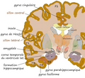
Coronal hippocampe.png 543 × 499; 298 KB
-
Coronal insula.png 560 × 409; 147 KB
-
CorpusCallosum.svg 1 025 × 598; 25 KB
-
Cunningham cerebral sulci.png 1 252 × 790; 2,84 MB
-
Desikan-Killiany atlas regions.pdf 3 150 × 1 918; 13,59 MB
-
Destrieux atlas regions.pdf 5 864 × 2 850; 28,77 MB
-
EB1911 Brain Fig. 11-Gyri and Sulci on Mesial aspect.jpg 682 × 393; 45 KB
-
EB1911 Brain Fig. 9-Gyri and Sulci.jpg 672 × 412; 57 KB
-
F. Vicq d'Azyr, Planches pour le traite de Wellcome L0000998.jpg 3 322 × 3 178; 5,03 MB
-
Face sup T1.png 563 × 482; 222 KB
-
Facies ventralis cerebri.jpg 1 834 × 2 068; 629 KB
-
File-Cerebral Gyri - Inferior Surface1.png 761 × 569; 419 KB
-
Frontal gyrus coronal sections.gif 148 × 158; 1,24 MB
-
Frontal gyrus sagittal sections.gif 185 × 158; 1,41 MB
-
Frontal gyrus transversal sections.gif 148 × 185; 1,39 MB
-
FrontalCapts.png 2 309 × 911; 927 KB
-
Gehirn Frontalschnitt hippocampus-it.png 1 055 × 573; 247 KB
-
Gehirn Frontalschnitt hippocampus.png 913 × 573; 263 KB
-
Gehirn lateral gyri el.svg 669 × 519; 67 KB
-
Gehirn lobi medial.png 819 × 598; 61 KB
-
Gehirn, lateral - Hauptgyri + Hauptsulci.svg 624 × 498; 56 KB
-
Gehirn, lateral - Hauptgyri beschriftet.svg 624 × 498; 334 KB
-
Gray1197.png 444 × 500; 47 KB
-
Gray725 anterior central gyrus.png 255 × 600; 61 KB
-
Gray725 cant been seen top.png 663 × 1 560; 205 KB
-
Gray725 frontal pole.png 255 × 600; 57 KB
-
Gray725 inferior frontal gyrus.png 255 × 600; 60 KB
-
Gray725 middle frontal gyrus.png 255 × 600; 61 KB
-
Gray725 occipital pole.png 255 × 600; 57 KB
-
Gray725 posterior central gyrus.png 255 × 600; 61 KB
-
Gray725 superior frontal gyrus.png 255 × 600; 61 KB
-
Gray725 superior parietal lobule.png 255 × 600; 60 KB
-
Gray725.png 255 × 600; 18 KB
-
Gray726 angular gyrus.png 992 × 573; 178 KB
-
Gray726 ar.svg 992 × 573; 409 KB
-
Gray726 cant been seen lateral.png 842 × 487; 177 KB
-
Gray726 frontal pole.png 992 × 573; 174 KB
-
Gray726 inferior frontal gyrus.png 992 × 573; 177 KB
-
Gray726 inferior parietal lobule (hy).png 2 108 × 1 217; 909 KB
-
Gray726 inferior parietal lobule.png 992 × 573; 182 KB
-
Gray726 inferior temporal gyrus.png 992 × 573; 169 KB
-
Gray726 middle frontal gyrus.png 992 × 573; 192 KB
-
Gray726 middle temporal gyrus.png 992 × 573; 173 KB
-
Gray726 occipital pole.png 992 × 573; 170 KB
-
Gray726 opecular part of IFG.png 992 × 573; 172 KB
-
Gray726 orbital part of IFG.png 992 × 573; 170 KB
-
Gray726 postcentral gyrus.png 992 × 573; 117 KB
-
Gray726 precentral gyrus.png 992 × 573; 181 KB
-
Gray726 superior frontal gyrus.png 992 × 573; 179 KB
-
Gray726 superior parietal lobule.png 992 × 573; 174 KB
-
Gray726 superior temporal gyrus.png 992 × 573; 173 KB
-
Gray726 supramarginal gyrus.png 992 × 573; 182 KB
-
Gray726 temporal pole.png 992 × 573; 170 KB
-
Gray726 triangular part of IFG.png 992 × 573; 174 KB
-
Gray726.png 700 × 405; 29 KB
-
Gray726.svg 992 × 573; 146 KB
-
Gray727 anterior cingulate cortex.png 1 025 × 598; 141 KB
-
Gray727 cant been seen medial.png 870 × 507; 147 KB
-
Gray727 cingulate gyrus.png 1 025 × 598; 92 KB
-
Gray727 Cuneus.png 1 025 × 598; 144 KB
-
Gray727 frontal pole.png 1 025 × 598; 137 KB
-
Gray727 fusiform gyrus.png 1 025 × 598; 135 KB
-
Gray727 inferior temporal gyrus.png 1 025 × 598; 134 KB
-
Gray727 latin.svg 1 025 × 598; 23 KB
-
Gray727 lingual gyrus.png 1 025 × 598; 134 KB
-
Gray727 occipital pole.png 1 025 × 598; 134 KB
-
Gray727 paracentral gyrus.png 1 025 × 598; 136 KB
-
Gray727 parahippocampal gyrus.png 1 025 × 598; 134 KB
-
Gray727 precuneus.png 1 025 × 598; 143 KB
-
Gray727 superior frontal gyrus.png 1 025 × 598; 142 KB
-
Gray727 temporal pole.png 1 025 × 598; 134 KB
-
Gray727 uncus of parahippocampal gyrus.png 1 025 × 598; 133 KB
-
Gray727.svg 1 025 × 598; 18 KB
-
Gray729 frontal pole.png 300 × 366; 60 KB
-
Gray729 orbital gyrus.png 300 × 366; 59 KB
-
Gray729 straight gyrus.png 300 × 366; 61 KB
-
Gray729.png 300 × 366; 20 KB
-
Gray743 cingulate gyrus.png 450 × 542; 60 KB
-
Gray743 inferior frontal gyrus.png 450 × 542; 172 KB
-
Gray743 insular cortex.png 450 × 542; 170 KB
-
Gray743 middle frontal gyrus.png 450 × 542; 171 KB
-
Gray743 straight gyrus.png 450 × 542; 170 KB
-
Gray743 superior frontal gyrus.png 450 × 542; 171 KB
-
Gray756.png 600 × 354; 32 KB
-
Gray757.png 600 × 346; 27 KB
-
Gyri and sulci of frontal cortex of monkey brain (Cebus apella).jpg 1 200 × 592; 99 KB
-
Gyri Basal no text1.png 572 × 719; 595 KB
-
Gyri Basal no text2.png 567 × 690; 584 KB
-
Gyri Insula no text.png 697 × 524; 399 KB
-
Gyri Lateral no text.png 632 × 484; 413 KB
-
Gyri Medial no text.png 644 × 498; 552 KB
-
Gyri Medial no text2.png 647 × 505; 444 KB
-
Gyrus cinguli.png 800 × 467; 116 KB
-
Gyrus externe droit.png 1 100 × 800; 261 KB
-
Gyrus externe.png 666 × 450; 112 KB
-
Gyrus sulcus ja.png 960 × 720; 115 KB
-
Gyrus sulcus.png 960 × 720; 95 KB
-
Hand-book of physiology (1892) (14578661130).jpg 1 796 × 1 516; 282 KB
-
Hand-book of physiology (1892) (14578725808).jpg 1 164 × 1 476; 185 KB
-
Hand-book of physiology (1892) (14785228633).jpg 1 776 × 1 128; 204 KB
-
Hippocampe parahippo.png 1 000 × 800; 426 KB
-
Homunculus-ja paracentral lobule.png 1 125 × 578; 189 KB
-
Human and chimp brain.png 1 010 × 1 346; 836 KB
-
Human brain inferior view description 2.JPG 373 × 467; 36 KB
-
Human brain inferior-medial view description 2.JPG 702 × 491; 68 KB
-
Human brain inferior-medial view description 3.JPG 702 × 491; 63 KB
-
Human brain inferior-medial view with marked Precuneus.JPG 702 × 491; 211 KB
-
Human brainstem anterior view 2 description.JPG 346 × 487; 35 KB
-
Human Cortical Development.png 2 303 × 2 656; 1 022 KB
-
Inferieur gyrus.png 724 × 652; 207 KB
-
Inferior frontal gyrus animation small.gif 150 × 150; 570 KB
-
Inferior frontal gyrus.png 300 × 190; 40 KB
-
Infero interne gyrus.png 579 × 449; 106 KB
-
Lateral surface - Inferior frontal gyrus.png 1 236 × 800; 597 KB
-
Lateral surface - Middle frontal gyrus.png 1 236 × 800; 596 KB
-
Lateral surface - opercular part of the inferior frontal gyrus.png 1 236 × 800; 593 KB
-
Lateral surface - Orbital part of inferior frontal gyrus.png 1 236 × 800; 594 KB
-
Lateral surface - Superior frontal gyrus.png 1 236 × 800; 594 KB
-
Lateral surface - triangular part of inferior frontal gyrus.png 1 236 × 800; 593 KB
-
Lateral surface of cerebral cortex - gyri.png 1 236 × 800; 760 KB
-
Lissencephaly.jpg 490 × 315; 17 KB
-
Lk444.jpg 500 × 667; 70 KB
-
Macaque monkey's premotor areas.jpg 1 200 × 1 108; 156 KB
-
Man&chimpbrains.png 591 × 558; 455 KB
-
Medial paracentral lob.png 673 × 481; 130 KB
-
Medial surface - Sperior frontal gyrus.png 1 179 × 747; 515 KB
-
Medial surface of cerebral cortex - ceneus.png 1 179 × 747; 514 KB
-
Medial surface of cerebral cortex - entorhinal cortex.png 1 179 × 747; 515 KB
-
Medial surface of cerebral cortex - fusiform gyrus.png 1 179 × 747; 515 KB
-
Medial surface of cerebral cortex - gyri.png 1 179 × 747; 644 KB
-
Medial surface of cerebral cortex - lingual gyrus.png 1 179 × 747; 515 KB
-
Medial surface of cerebral cortex - parahippocampal.png 1 179 × 747; 515 KB
-
Medial surface of cerebral cortex - preceneus.png 1 179 × 747; 514 KB
-
Middle frontal gyrus.png 300 × 190; 42 KB
-
OccCapts.png 1 662 × 781; 847 KB
-
Operculum.png 700 × 405; 59 KB
-
Operculum1.jpg 1 065 × 796; 146 KB
-
Orbital gyrus viewed from bottom.png 504 × 637; 268 KB
-
Paracentral lobule animation small.gif 150 × 150; 568 KB
-
Parahippocampe.png 1 032 × 761; 622 KB
-
ParcellationBrains.jpg 364 × 384; 118 KB
-
ParietCapts.png 2 337 × 878; 819 KB
-
Postcentral gyrus 3d.png 600 × 600; 177 KB
-
Postcentral gyrus.gif 250 × 250; 1,87 MB
-
Pre- and post-central gyrus, right hemisphere cropped.png 426 × 488; 140 KB
-
Pre- and post-central gyrus, right hemisphere.jpg 884 × 1 394; 1,13 MB
-
Precentral gyrus 3d.png 600 × 600; 178 KB
-
Precentral gyrus.jpg 700 × 405; 47 KB
-
Precuneus connectivity new.gif 343 × 447; 30 KB
-
Precuneus connectivity.jpg 4 111 × 6 939; 81,65 MB
-
PretermSurfaces HiRes es.png 876 × 190; 158 KB
-
PretermSurfaces HiRes ja.png 876 × 190; 146 KB
-
PretermSurfaces HiRes.png 876 × 190; 165 KB
-
PSM V35 D759 Diagram of the left cerebral hemisphere.jpg 1 626 × 1 413; 254 KB
-
Sagittale-insula-Heschl.png 579 × 420; 140 KB
-
Slide2HAN.JPG 960 × 720; 96 KB
-
Slide3HAN.JPG 960 × 720; 117 KB
-
Smith.PNG 1 500 × 928; 735 KB
-
Sobo 1909 623 ar.png 2 404 × 2 652; 5,62 MB
-
Sobo 1909 623.png 2 404 × 2 652; 18,27 MB
-
Sobo 1909 624.png 3 060 × 2 247; 19,71 MB
-
Sobo 1909 625.png 1 137 × 703; 2,29 MB
-
Sobo 1909 626.png 1 326 × 899; 3,42 MB
-
Sobo 1909 627.png 983 × 981; 2,76 MB
-
Sobo 1909 628.png 1 058 × 1 159; 3,52 MB
-
Sobo 1909 629.png 991 × 1 015; 98 KB
-
Sobo 1909 630.png 1 077 × 1 239; 3,83 MB
-
Sobo 1909 631.png 1 010 × 689; 1,99 MB
-
Sobo 1909 632.png 1 363 × 879; 3,43 MB
-
Sobo 1909 633.png 1 078 × 699; 2,16 MB
-
Sobo 1909 634.png 1 089 × 703; 2,19 MB
-
Some brain areas.png 989 × 412; 146 KB
-
Standard anatomical parcellation of the posterior cortical surface.png 1 810 × 1 717; 1,26 MB
-
Straight gyrus - inferior view.png 500 × 638; 284 KB
-
Straight gyrus animation.gif 300 × 300; 1,81 MB
-
Superieur gyrus.png 554 × 516; 115 KB
-
Superior frontal gyrus animation small.gif 150 × 150; 549 KB
-
Superior frontal gyrus.png 300 × 190; 32 KB
-
Superior temporal gyrus.png 300 × 190; 35 KB


