Category:Hip dysplasia
Zur Navigation springen
Zur Suche springen
Fehlstellung des Hüftgelenks | |||||
| Medium hochladen | |||||
| Ist ein(e) | |||||
|---|---|---|---|---|---|
| Unterklasse von |
| ||||
| |||||
Unterkategorien
Es werden 5 von insgesamt 5 Unterkategorien in dieser Kategorie angezeigt:
In Klammern die Anzahl der enthaltenen Kategorien (K), Seiten (S), Dateien (D)
A
H
X
Medien in der Kategorie „Hip dysplasia“
Folgende 48 Dateien sind in dieser Kategorie, von 48 insgesamt.
-
 4montholdwearingfrejkabracetocorrecthipdysplasia.jpg 604 × 454; 31 KB
-
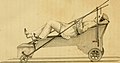
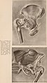
 A treatise on orthopedic surgery (1910) (14783095002).jpg 1.398 × 1.298; 397 KB
-
-
 Aceetabuloplastik (Pemberton osteotomy).jpg 2.186 × 1.700; 334 KB
-
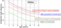
 Acetabular index by age in females.png 2.050 × 1.134; 215 KB
-
 Acetabular index by age in females.svg 1.537 × 850; 28 KB
-
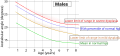
 Acetabular index by age in males.png 2.070 × 956; 204 KB
-
 Acetabular index by age in males.svg 1.553 × 717; 32 KB
-
 Acetabulopl. nach Keilinterposition.jpg 377 × 661; 19 KB
-
 Acetabuloplastik mit Keil.png 200 × 165; 7 KB
-
 Acetabuloplastik OP Schritt 2.jpg 207 × 153; 6 KB
-
 Acetabuloplastik-Schritt1.svg 640 × 520; 14 KB
-
 Acetabuloplastik-Schritt2.svg 640 × 520; 14 KB
-
 Acetabuloplastik-Schritt3.svg 600 × 520; 14 KB
-
 Acetabuloplastik-Schritt4.svg 600 × 520; 14 KB
-
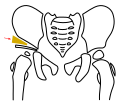
 Biomechanik-Hueftdysplasie.png 640 × 486; 26 KB
-
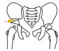
 Biomechanik-Hüftdysplasie-dysplastisch.svg 322 × 338; 5 KB
-
 Biomechanik-Hüftdysplasie-Normalstellung.svg 322 × 338; 5 KB
-
 Congenital dislocation of the hip Wellcome L0061332.jpg 3.354 × 5.646; 2,83 MB
-
 Congenital dislocation of the hip Wellcome L0062609.jpg 3.437 × 5.710; 3,02 MB
-
 DBO prä op CT-VRT.jpg 420 × 341; 19 KB
-
 Frejka.jpg 363 × 499; 29 KB
-
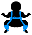
 Frejka.svg 626 × 696; 13 KB
-
 Hip dysplasia - schematic.jpg 4.497 × 1.476; 342 KB
-
 Hip dysplasia ultrasound.svg 725 × 510; 9 KB
-
 Hüftdysplasie Skizze Basis.svg 750 × 650; 16 KB
-
 Image quality checking of pediatric pelvis.jpg 1.040 × 569; 201 KB
-
 Interstate medical journal (1917) (14597230797).jpg 1.508 × 1.608; 225 KB
-
 Interstate medical journal (1917) (14780566771).jpg 2.248 × 2.810; 374 KB
-
 Interstate medical journal (1917) (14783364172).jpg 1.826 × 3.256; 609 KB
-
 Interstate medical journal (1917) (14803561673).jpg 2.110 × 3.062; 499 KB
-
 Klasyfikacja Grafa - kąty alfa i beta w stawie biodrowym.jpg 725 × 616; 27 KB
-
 Living anatomy and pathology; (1910) (14571489010).jpg 2.212 × 3.188; 578 KB
-
 Pavlik.jpg 336 × 497; 32 KB
-
 Pavlik.svg 626 × 696; 16 KB
-
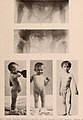
 Pediatrics. (1897) (14596748528).jpg 1.422 × 2.258; 429 KB
-
 Pemberton-pelvic-osteotomy step1.jpg 2.032 × 1.510; 243 KB
-
 Pemberton-pelvic-osteotomy step2.jpg 2.032 × 1.510; 251 KB
-
 Pemberton-pelvic-osteotomy step3.jpg 2.032 × 1.510; 243 KB
-
 Pemberton-pelvic-osteotomy step4.jpg 1.949 × 1.515; 243 KB
-
 Saeugling mit angelegter spreizhose.jpg 2.000 × 1.500; 876 KB
-
 Salter-Osteotomie-Schritt1.svg 640 × 520; 14 KB
-
 Salter-Osteotomie-Schritt2.svg 600 × 520; 13 KB
-
 Salter-Osteotomie-Schritt3.svg 600 × 520; 14 KB
-
 Salter-Osteotomie-Schritt4.svg 570 × 520; 13 KB
-
 Salter-Osteotomie.gif 200 × 155; 5 KB
-
 Tractie.jpg 698 × 701; 137 KB
-
 Tübinger Schiene.jpg 409 × 417; 44 KB

_(14783095002).jpg)
_(14590014809).jpg)
.jpg)


_(14597230797).jpg)
_(14780566771).jpg)
_(14783364172).jpg)
_(14803561673).jpg)

_(14571489010).jpg)


_(14596748528).jpg)


