Category:Insular cortex
Zur Navigation springen
Zur Suche springen
eingesenkter Teil der Großhirnrinde | |||||
| Medium hochladen | |||||
| Ist ein(e) |
| ||||
|---|---|---|---|---|---|
| Unterklasse von |
| ||||
| Ist Teil von | |||||
| Verschieden von | |||||
| |||||
Unterkategorien
Es werden 2 von insgesamt 2 Unterkategorien in dieser Kategorie angezeigt:
In Klammern die Anzahl der enthaltenen Kategorien (K), Seiten (S), Dateien (D)
C
- Central insular sulcus (6 D)
- Circular sulcus of insula (10 D)
Medien in der Kategorie „Insular cortex“
Folgende 55 Dateien sind in dieser Kategorie, von 55 insgesamt.
-
"Traite complet de l'anatomie...",Foville, 1844 Wellcome L0019131.jpg 1.215 × 1.561; 418 KB
-
Anatomy of insula.jpg 612 × 366; 197 KB
-
Areas characterized by the presence of VENs in the human brain.jpg 892 × 630; 72 KB
-
Brain areas that participate in social processing.jpg 828 × 308; 193 KB
-
Brain lobes - insular lobe.png 642 × 482; 406 KB
-
Brain model - Insula.jpg 640 × 480; 73 KB
-
Brodmann areas inside of lateral sulcus close up.png 772 × 637; 211 KB
-
Brodmann areas inside of lateral sulcus.png 792 × 839; 1,91 MB
-
Cerebral Gyri - Insula.png 761 × 571; 293 KB
-
Classification and dissection of the insula.png 1.935 × 1.275; 2,38 MB
-
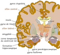
Coronal hippocampe.png 543 × 499; 298 KB
-
Error-related activations during a stop signal task.jpg 512 × 458; 163 KB
-
Gray658.png 361 × 450; 40 KB
-
Gray717-emphasizing-insula.png 500 × 568; 294 KB
-
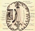
Gray731.png 600 × 342; 64 KB
-
Gray743 insular cortex.png 450 × 542; 170 KB
-
Human and chimp brain.png 1.010 × 1.346; 836 KB
-
Human brain frontal (coronal) section description2.JPG 702 × 487; 42 KB
-
Human brain view on transverse temporal and insular gyri.JPG 470 × 326; 22 KB
-
Human Insular Anatomy.png 858 × 546; 361 KB
-
Inflated surface of the left hemisphere and insular region.png 861 × 319; 330 KB
-
Insula animation small.gif 150 × 150; 598 KB
-
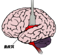
Insula cortex ja.png 700 × 645; 128 KB
-
Insula of human and cynomolgus macaque monkey.png 2.361 × 1.694; 3,07 MB
-
Insula structure.png 650 × 364; 164 KB
-
Insular cortex and connecting brain regions.png 675 × 372; 49 KB
-
Insular cortex coronal sections.gif 148 × 158; 1,2 MB
-
Insular cortex granulation.png 726 × 400; 38 KB
-
Insular cortex of human brain (left hemisphere).jpg 960 × 720; 44 KB
-
Insular cortex sagittal sections.gif 185 × 158; 1,19 MB
-
Insular Cortex Sub-regions.png 1.299 × 655; 448 KB
-
Insular cortex transversal sections.gif 148 × 185; 1,23 MB
-
Insular cortex.gif 500 × 390; 8,86 MB
-
Lawrence 1960 2.21-23.png 2.024 × 2.840; 1,58 MB
-
Lawrence 1960 2.3.png 1.616 × 952; 578 KB
-
Operculum.png 700 × 405; 59 KB
-
Operculum1.jpg 1.065 × 796; 146 KB
-
PretermSurfaces HiRes es.png 876 × 190; 158 KB
-
PretermSurfaces HiRes ja.png 876 × 190; 146 KB
-
PretermSurfaces HiRes.png 876 × 190; 165 KB
-
Recompenses.jpg 619 × 347; 51 KB
-
Regeczy780.jpg 1.308 × 1.167; 158 KB
-
Sagittale-insula-Heschl.png 579 × 420; 140 KB
-
Smoking insula.jpg 600 × 450; 31 KB
-
Sobo 1909 633.png 1.078 × 699; 2,16 MB
-
Sobo 1909 635.png 1.045 × 1.043; 3,12 MB
-
Sobo 1909 642.png 1.063 × 1.019; 3,11 MB
-
Sobo 1909 645.png 1.227 × 750; 2,64 MB
-
Sobo 1911 644.png 2.496 × 1.692; 12,1 MB
-
The insula and connecting brain regions.png 1.696 × 996; 400 KB
-
Three anterior short gyri.png 415 × 333; 76 KB
-
Two-and three-dimensional images of the anterior and posterior insular cortices.png 4.051 × 3.329; 2,25 MB
-
Vies interoceptives.png 2.470 × 1.138; 1,48 MB


