Category:Laboratory techniques
Vai alla navigazione
Vai alla ricerca
| Carica un file multimediale | |||||
| Sottoclasse di |
| ||||
|---|---|---|---|---|---|
| |||||
Sottocategorie
Questa categoria contiene le 32 sottocategorie indicate di seguito, su un totale di 32.
*
A
B
C
- Cannula transfer (7 F)
E
F
- Franz cells (1 F)
- Function-spacer-lipid construct (13 F)
G
I
- Isotope dilution (8 F)
L
M
- Murashige and Skoog medium (5 F)
P
- Patch-clamp techniques (120 F)
S
V
- Vargulin (2 F)
File nella categoria "Laboratory techniques"
Questa categoria contiene 174 file, indicati di seguito, su un totale di 174.
-
-

-
 201019-N-DA693-1002.jpg 4 044 × 2 889; 1,12 MB
-
 505 Figure.png 5 500 × 800; 599 KB
-
 6870 Freezer Mill.jpg 3 264 × 2 176; 2,47 MB
-
 AAV Gene Therapy.jpg 495 × 371; 137 KB
-
 Ajouter réactifs 1.png 309 × 514; 10 KB
-
 Ajouter réactifs 2.png 309 × 514; 10 KB
-
 Aliquoting after spinning down.jpg 2 826 × 2 574; 1 005 KB
-
 AN-VI Hysteresis.jpg 1 207 × 419; 172 KB
-
 AN-VI isteresi.JPG 1 207 × 419; 68 KB
-
 AN-VI Transitorio accelerazione.png 675 × 424; 26 KB
-
 AN-VI Transitorio spostamento.png 448 × 419; 24 KB
-
 AN-VI Transitorio.png 441 × 427; 20 KB
-
 Anthrax culture.jpg 1 050 × 700; 254 KB
-
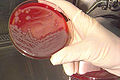
 Antonio e Biagio e Cesare Arrigo Emocoltura.jpg 3 264 × 2 448; 4,55 MB
-
 Apoptotic DNA Laddering.png 178 × 193; 20 KB
-
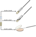 Aseptic transfer from broth.png 958 × 984; 91 KB
-
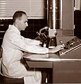
 Assayer, 1916.jpg 845 × 1 208; 260 KB
-
 AutoFISH.jpg 1 348 × 1 180; 1,33 MB
-
 BAC work flow ESP.jpg 1 234 × 1 113; 75 KB
-
 Balizas moleculares.png 960 × 720; 11 KB
-
 Band vs Boundary Ultracentrifugation.svg 662 × 444; 20 KB
-
 Bestimmung der Rohdichte von RSS Flüssigboden®.jpg 200 × 267; 8 KB
-
 Biolumj.JPG 1 042 × 538; 147 KB
-
 Body fluid cell count testing on automated analyzer.jpg 2 825 × 3 489; 1,86 MB
-
 Boom method by Tajima pipet.jpg 942 × 400; 96 KB
-
 Calcímetro de Bernard.jpg 713 × 671; 79 KB
-
 Capillary blot setup.svg 307 × 123; 43 KB
-
 ChIPPET3.JPG 720 × 540; 24 KB
-
 ChIPPET4.JPG 720 × 540; 16 KB
-
 ChIPPET5.JPG 720 × 540; 19 KB
-
 ChIPPET6.JPG 720 × 540; 13 KB
-
 ChIPPET7.JPG 720 × 540; 13 KB
-
 ChIPPET8.JPG 720 × 540; 32 KB
-
 ChIPPET9.JPG 720 × 540; 26 KB
-
 Coagulase test in S. aureus and S. epidermidis.jpg 723 × 300; 160 KB
-
 Coagulase+.JPG 574 × 744; 289 KB
-
 Courtney 2008.jpg 786 × 685; 52 KB
-
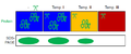
 Cut-offs of different liquid filtration techniques.png 1 200 × 900; 3,09 MB
-
 Cytoxydase.jpg 564 × 300; 143 KB
-
 Deconvoluted ESMS.jpg 1 648 × 1 123; 71 KB
-
 Demonstration of Method- Osmium-μCT with Advanced Image Processing.tif 4 683 × 1 996; 35,66 MB
-
 Destilační aparatura.webp 1 000 × 1 338; 124 KB
-
 Determinación de Fe y Zn en muestras de músculo de ternera.jpg 3 366 × 3 024; 4,81 MB
-
 Distillation 2-2.jpg 2 592 × 1 710; 1,63 MB
-
 Distillation 2-3.jpg 2 544 × 1 509; 1,44 MB
-
 Distillation 2-a.jpg 2 592 × 1 944; 1,71 MB
-
 Distillation 2-b.jpg 2 592 × 1 944; 2,1 MB
-
 Distillation 2.jpg 2 592 × 1 944; 1,3 MB
-
 Distillation setup 2.JPG 3 264 × 4 912; 2,88 MB
-
 Distillation setup.JPG 3 264 × 4 912; 2,8 MB
-

-
 Dělicí nálevka ve filtračním kruhu.jpg 1 200 × 1 600; 116 KB
-
 Dělicí nálevka.webp 1 000 × 1 338; 73 KB
-
-
 Elasticità di rimbalzo.jpg 385 × 503; 131 KB
-

-
 ELISA diretto e sandwich.png 700 × 700; 99 KB
-
 Exclusion cells.jpg 2 500 × 1 666; 1,41 MB
-
 Extrakční aparatura.jpg 1 200 × 1 600; 136 KB
-
 FASTpp cartoon.png 481 × 200; 18 KB
-
 Fatma Sri Wahyuni 3.jpg 588 × 960; 51 KB
-
 FernadezMoran.jpg 230 × 239; 15 KB
-
 Forschungsschwerpunkt Biotechnologie.jpg 4 288 × 2 848; 6,55 MB
-
 FRET cz CFP YFP.tif 602 × 426; 57 KB
-
 FRET cz jablonskeho diagram.tif 528 × 351; 22 KB
-
 FRET cz kappa2 orientation factor.tif 1 290 × 1 426; 299 KB
-
 FRET cz photobleaching.tif 649 × 462; 278 KB
-
 FRET cz proteolyza.tif 338 × 811; 202 KB
-
 FRET cz zavislost ucinnosti na ruznych faktorech.tif 636 × 914; 1,67 MB
-
 GCruleof10.jpg 450 × 300; 27 KB
-
 Gomma Transitorio accelerazione.png 785 × 492; 71 KB
-
 Gomma Transitorio spostamento.png 446 × 421; 40 KB
-
 Grafica de la PCR.jpg 528 × 600; 77 KB
-
 Grafico loss factor polito.jpg 558 × 593; 136 KB
-
 Gélose Rambach.JPG 2 272 × 1 704; 1,79 MB
-
 Hibridación in situ fluorescente (FISH del gen ALK).jpg 1 920 × 1 037; 102 KB
-
 Hydroxy-probing.png 183 × 214; 29 KB
-
 IMSI análisis del espermatozoïde a más de 6000X.jpg 396 × 306; 8 KB
-
 IMSI espermatozoides en gota de selección.jpg 514 × 396; 16 KB
-
 IMSI inmovilización del espermatozoide.jpg 466 × 375; 8 KB
-

-
 In solution capture.png 630 × 655; 46 KB
-
 Indole.PNG 696 × 157; 14 KB
-
 Indole.svg 668 × 129; 34 KB
-
 Jodometrická titrace 2.jpg 719 × 1 280; 93 KB
-
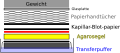 Kapillarblot.svg 282 × 123; 42 KB
-
 Kapkovací destička.jpg 1 152 × 2 048; 79 KB
-
 Knockoutmouse80-72.jpg 772 × 401; 42 KB
-
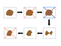 Kvartace-plain.png 720 × 540; 5 KB
-
 Kvartace-čísla.png 720 × 540; 5 KB
-
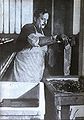
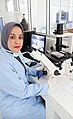
 Lab technician- Lady.jpg 2 136 × 2 956; 1,37 MB
-
 Lab thermometer.JPG 3 264 × 4 912; 2,29 MB
-
 LAMP method.jpg 300 × 300; 48 KB
-
 Liebig condensation.JPG 4 912 × 3 264; 3,31 MB
-
 Load a sample into a agarose gel.jpg 3 456 × 2 304; 2,17 MB
-
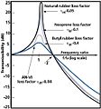 Loss factor eng polito.jpg 556 × 596; 41 KB
-
 Mason Weaver cell.png 458 × 202; 4 KB
-
 Medical Support Squadron provide for Icemen 111115-F-HA566-036.jpg 4 256 × 2 832; 4,42 MB
-
 Michaelis-Menten plot.svg 1 020 × 641; 104 KB
-
 Mixing the medications at the laminar flow cabinet.jpg 3 072 × 2 048; 352 KB
-
 MRI method copy.tif 4 047 × 2 313; 35,71 MB
-
 MTT Plate.jpg 1 024 × 768; 250 KB
-
 Méthode de Wijs schema du dosage.jpg 400 × 294; 9 KB
-
 NASA Goddard technologist studies a paint sample in her laboratory.jpg 1 000 × 1 115; 128 KB
-
 NHGRI researcher uses a pipette to remove DNA from a micro test tube.jpg 3 008 × 1 960; 1,72 MB
-
 Northern Blot Scheme.PNG 640 × 400; 14 KB
-
 Nuč s fritou v odsávací aparatuře.webp 422 × 750; 32 KB
-
 Odsávací aparatura.jpg 6 936 × 9 248; 10,81 MB
-
Otevírání ampule bromu.webm 1 min 14 s, 1 920 × 1 080; 25,25 MB
-
 Parik1.jpg 680 × 552; 65 KB
-
 Parik2.1.jpeg 388 × 636; 29 KB
-
 Parik3.jpeg 623 × 642; 70 KB
-
 Parik4.jpg 706 × 334; 55 KB
-
 Passiflora micropropagation Annalisa Giovannini DSCN1999.jpg 3 456 × 4 608; 3,46 MB
-
 Pcts image.jpg 788 × 616; 311 KB
-
 Peroxide Fluxer from Claisse Corporation.jpg 316 × 273; 22 KB
-
 PET deletion insertion.PNG 685 × 613; 64 KB
-
 PETworkflow.png 1 061 × 797; 75 KB
-
 Pexeso filtrační aparatura pro filtraci za sníženého tlaku 1.jpg 9 280 × 6 944; 11,41 MB
-
 Pexeso filtrační aparatura pro filtraci za sníženého tlaku 2.jpg 9 280 × 6 944; 11,99 MB
-
 Pipette Graph.jpg 1 596 × 813; 58 KB
-
 Precipitación-salina-huevo.jpg 2 928 × 1 744; 1,52 MB
-
 Precision cut urine slices.jpg 708 × 320; 142 KB
-
 Preparation of the modified cellulose-based complex particle.jpg 610 × 496; 86 KB
-

-
 Produkt rekrystalizace.jpg 1 200 × 1 600; 80 KB
-
 Proves de la TaqMan.jpg 650 × 167; 23 KB
-
 Průběh rekrystalizace.jpg 1 200 × 1 600; 115 KB
-
 Průběh syntézy chalkonu.jpg 6 944 × 9 280; 10,59 MB
-
 Přidávání látky do reakční směsi pomocí skleněné Pasteurovy pipety.jpg 1 200 × 1 600; 151 KB
-
 Přikapávací nálevka v aparatuře.webp 1 000 × 1 338; 129 KB
-
 R loop.jpg 720 × 540; 72 KB
-
 R SIVA SUBRAMNIAN PRACTICAL ASSIGNMENT PG DIMLOMA IN HOSPITAL LABORATORY TECHNOLOGY.pdf 1 275 × 1 650, 22 pagine; 1,05 MB
-
 Rbound elasticity.jpg 385 × 503; 131 KB
-
 Rebound.JPG 395 × 555; 41 KB
-
 Recta de calibratge de la PCR.png 800 × 308; 65 KB
-
 Reticulo Neubauer.jpg 836 × 571; 82 KB
-
 Rheometer-brookfield-rsx.png 1 170 × 2 061; 2,67 MB
-
 Rimbalzo.jpg 1 354 × 1 689; 141 KB
-
 RNA agarose gel.svg 728 × 732; 198 KB
-
 RNA-PET.PNG 1 066 × 807; 46 KB
-
 Rosiglitazone and Exercise Effect on MAT.pdf 585 × 247; 642 KB
-
 Rosiglitazone and Exercise Effect on MAT.svg 800 × 344; 624 KB
-
 Rychlá filtrace.jpg 422 × 750; 23 KB
-
 Schéma aparatury filtrace za sníženého tlaku.png 250 × 143; 19 KB
-
 ScorpionsProbes.jpg 640 × 480; 60 KB
-
 Seal f film hidrofobic property.jpg 1 480 × 740; 429 KB
-
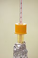
 Siwoloboff method.png 542 × 802; 17 KB
-
 Sondes balisa.jpg 220 × 165; 34 KB
-
 Sondes scorpion.jpg 220 × 165; 37 KB
-
 Sonicator.jpg 2 448 × 3 264; 3,14 MB
-
 Správné a chybné upevnění křížové svorky.png 365 × 228; 28 KB
-
 StandardAddition.png 683 × 467; 20 KB
-
 Stanovení obsahu cukru v mouce.jpg 1 536 × 2 048; 262 KB
-
 Sublimed camphor.jpg 416 × 413; 14 KB
-
 Sysmex WBC scattergrams.jpg 538 × 282; 25 KB
-
 Teen scientist Alexa Dantzler in the lab.jpg 2 292 × 1 719; 394 KB
-
 Titrace za horka.jpg 422 × 750; 24 KB
-
 Todd synthesis.png 697 × 196; 5 KB
-
 Tombstone courthouse assay lab.jpg 2 145 × 1 950; 397 KB
-
 Trojhrdlá.webp 422 × 750; 40 KB
-
 Tube condenser.png 2 221 × 1 672; 2,6 MB
-

-
 Verdünnungsreihe mit Ausplattieren.svg 491 × 308; 2,49 MB
-
 Vintage students laboratory (1904) at Gdansk University of Technology, Poland.jpg 6 000 × 3 376; 4,88 MB
-
 Vintage students laboratory (1904), Gdansk University of Technology, Poland.jpg 6 000 × 3 376; 5 MB
-
 Vintage students laboratory room (1904), Gdansk University of Technology, Poland.jpg 6 000 × 3 376; 5,13 MB
-
 Virus counter.jpg 3 078 × 1 282; 1,83 MB
-
 Working Procedure for calibration.jpg 347 × 276; 20 KB
-
 Zone of Inhibition.jpg 1 152 × 864; 319 KB
-
 Świecenie luminolu w teście na obecność krwi, Centrum Nauki Kopernik, laboratorium.jpg 3 804 × 2 750; 5,45 MB
-
 Геолог в лаборатории.jpg 3 403 × 2 268; 1,01 MB

_%D0%A4%D0%BE%D1%82%D0%BE,_%D0%BE%D1%81%D0%B0%D0%B6%D0%B4%D0%B5%D0%BD%D0%BD%D0%BE%D0%B9_%D0%94%D0%9D%D0%9A_%D0%B2_96%25_%D1%81%D0%BF%D0%B8%D1%80%D1%82%D0%B5.jpg)


.jpg)


_at_Gdansk_University_of_Technology,_Poland.jpg)
,_Gdansk_University_of_Technology,_Poland.jpg)
,_Gdansk_University_of_Technology,_Poland.jpg)


