Category:Macrophages
Przejdź do nawigacji
Przejdź do wyszukiwania
Bahasa Indonesia: Makrofag
Bosanski: Makrofag
Català: Macròfag
Čeština: Makrofág
Deutsch: Makrophage
Español: Macrófago
Euskara: Makrofago
Français : Macrophage
Hrvatski: Makrofag
Italiano: Macrofago
Lietuvių: Makrofagas
Nederlands: Macrofaag
Polski: Makrofag
Português: Macrófago
Slovenčina: Makrofág
Suomi: Makrofagi
Svenska: Makrofag
Tiếng Việt: Đại thực bào
Türkçe: Makrofaj
Български: Макрофаг
Русский: Макрофаги
Српски / srpski: Макрофаг
Українська: Макрофаги
日本語: マクロファージ
中文: 巨噬细胞
עברית : מקרופאג
العربية : بلعم
فارسی : درشتخوار
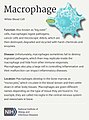
type of white blood cell  | |||||
| Prześlij plik multimedialny | |||||
| Jest to | |||||
|---|---|---|---|---|---|
| Podklasa dla |
| ||||
| Poprzednik | |||||
| |||||
Podkategorie
Poniżej wyświetlono 9 spośród wszystkich 9 podkategorii tej kategorii.
- SVG macrophages (9 plików)
?
- Alveolar macrophages (25 plików)
F
- Foam cell (4 pliki)
G
M
- Macrophage activation (18 plików)
- Macrophage polarization (4 pliki)
P
- Peritoneal macrophages (8 plików)
S
- Siderophages (2 pliki)
Pliki w kategorii „Macrophages”
Poniżej wyświetlono 68 spośród wszystkich 68 plików w tej kategorii.
-
Abklingphase einer Entzündung.webm 43 s, 1920 × 1080; 43,3 MB
-

-
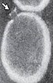
 African swine fever infected macrophage.jpg 640 × 432; 66 KB
-
 Anishkow cells.jpg 1278 × 684; 75 KB
-
 Autophagy in macrophage.jpg 836 × 836; 321 KB
-
 BMDM production.png 720 × 405; 39 KB
-
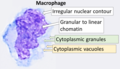 Cytology of a macrophage.png 1097 × 629; 511 KB
-
 Fimmu-09-02733-g001.jpg 963 × 748; 459 KB
-
 Fimmu-09-02733-g002.jpg 959 × 881; 548 KB
-
 From Immunity With Love.JPG 784 × 840; 56 KB
-
 Giemsa Stain Macrophage Illustration.png 2082 × 1807; 2,9 MB
-
 Gram stain of a macrophage with ingested S epidermidis bacteria.jpg 2048 × 1532; 326 KB
-
 Heme Breakdown ru.png 868 × 993; 95 KB
-
 Heme Breakdown.png 812 × 993; 86 KB
-
 Hemophagocytosis 1.jpg 397 × 294; 68 KB
-
 Hemophagocytosis 2.jpg 397 × 294; 22 KB
-

-

-
 Histopathology of a smoker's macrophage.jpg 549 × 529; 75 KB
-
 Histopathology of anthracotic macrophage in lung, annotated.jpg 354 × 349; 48 KB
-
 Histopathology of anthracotic macrophage in lung.jpg 354 × 349; 49 KB
-
 HIV on macrophage.png 1417 × 1417; 555 KB
-
 Human Cell Groups distributed by Cell Count and by Aggregate Cell Mass.jpg 3162 × 2096; 1,08 MB
-
 Leish amastig macrofago.jpg 3264 × 2448; 1,16 MB
-
 MAC 1.png 671 × 702; 559 KB
-
 MAC II.jpg 411 × 631; 19 KB
-
 Macrophage (17195150690).jpg 640 × 444; 115 KB
-
 Macrophage (30623015202).jpg 1633 × 2203; 470 KB
-
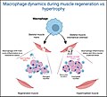 Macrophage dynamics during muscle regeneration vs. Hypertrophy.jpg 2128 × 1917; 322 KB
-
 Macrophage in the alveolus Lung - TEM.jpg 640 × 480; 150 KB
-
 Macrophage Polarization (M1 and M2 Macrophage).jpg 1084 × 566; 355 KB
-
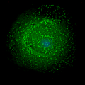
 Macrophage.jpg 1280 × 1024; 279 KB
-
 Macrophage.png 174 × 149; 17 KB
-
 Macrophages 001 (2575271746).jpg 430 × 446; 108 KB
-
 Macrophages 004 (2575258744).jpg 418 × 417; 99 KB
-
 Macrophages 01.jpg 640 × 382; 24 KB
-
 Macrophages 02.jpg 640 × 435; 80 KB
-
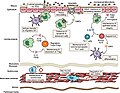
 Macrophages and helper T-cells.jpg 960 × 720; 49 KB
-
 Macrophages undergo mitosis after ingesting a fungal cell.jpg 2551 × 3300; 4,84 MB
-
 Macrófago (Macrophage) (35795300574).jpg 1176 × 1731; 572 KB
-
 Macs killing cancer cell.jpg 2289 × 1669; 1,08 MB
-
 MARCO Domain Structure.jpg 589 × 581; 45 KB
-
 Maturazione dei fagociti mononucleati.jpg 1515 × 449; 64 KB
-
 Melanin laden macrophages in dermatopathic lymphadenopathy 20X.jpg 816 × 664; 372 KB
-
 Melanin laden macrophages in dermatopathic lymphadenopathy 40X.jpg 411 × 311; 27 KB
-
 Melanin-laden macrophages in dermatopathic lymphadenopathy.jpg 710 × 663; 259 KB
-
 Melanophage.jpg 112 × 146; 17 KB
-
 Micrograph of a melanophage.jpg 379 × 331; 39 KB
-
 Microscopy of a bronchoalveolar lavage sample.jpg 823 × 268; 303 KB
-
 Mouse embryo Cellular expansion and morphology of CSF1R+ progenitors.jpg 1983 × 1946; 2,73 MB
-
 Mouse embryo Intravascular trafficking is independent of MYB and CX3CR1.jpg 1360 × 2512; 1,84 MB
-
 Mouse embryo Intravascular trafficking of CX3CR1+ YS pre-macrophages.jpg 1780 × 2460; 2,62 MB
-
 Mouse embryo Pre-macrophages infiltrate embryonic tissues.jpg 1346 × 2383; 2,14 MB
-
 Mouse embryo Trafficking is associated with cellular morphology.jpg 1578 × 1923; 1,16 MB
-
 Mouse embryo Trafficking kinetics of CSF1R+ cells are similar to pre-macrophages.jpg 1790 × 1508; 1,34 MB
-
 Mouse embryo Trafficking of KIT+ EMPs.jpg 1358 × 1888; 1,2 MB
-
 Opsonin.png 859 × 560; 108 KB
-
 PGC-1 alpha and Exercise cross-talk.jpg 1817 × 780; 1,89 MB
-
 Picture2 deutsch.jpg 478 × 799; 394 KB
-
 Picture2 englisch.jpg 478 × 501; 114 KB
-
-

-
 Series004Snapshot1 ch00.tif 1489 × 1489; 3,74 MB
-

-
 The effects of exercise on pro-inflammatory cytokines.png 850 × 621; 232 KB
-
 Tingible body macrophage.jpg 600 × 453; 212 KB
-
 Tumour Associated Macrophage (TAM).jpg 1080 × 968; 163 KB
-
 Yolk sac macrophage progenitors traffic to the embryo.png 685 × 438; 238 KB


.jpg)
.jpg)


.jpg)


.jpg)
.jpg)


_(35795300574).jpg)


.jpg)
