Category:Mesoderm
Zur Navigation springen
Zur Suche springen
mittleres Keimblatt | |||||
| Medium hochladen | |||||
| Ist ein(e) |
| ||||
|---|---|---|---|---|---|
| Unterklasse von |
| ||||
| |||||
Unterkategorien
Es werden 3 von insgesamt 3 Unterkategorien in dieser Kategorie angezeigt:
In Klammern die Anzahl der enthaltenen Kategorien (K), Seiten (S), Dateien (D)
Medien in der Kategorie „Mesoderm“
Folgende 200 Dateien sind in dieser Kategorie, von 227 insgesamt.
(vorherige Seite) (nächste Seite)-
 41392 2022 1024 Fig1 HTML.webp 1.799 × 1.357; 128 KB
-
 A human embryo of 2 mm. in median sagittal section.jpg 838 × 1.006; 310 KB
-

-

-
A-Comprehensive-Panel-of-Three-Dimensional-Models-for-Studies-of-Prostate-Cancer-Growth-Invasion-pone.0010431.s013.ogv 1 min 6 s, 524 × 384; 8,71 MB
-
A-Multi-cell-Multi-scale-Model-of-Vertebrate-Segmentation-and-Somite-Formation-pcbi.1002155.s016.ogv 1 min 3 s, 640 × 480; 6,71 MB
-
A-Multi-cell-Multi-scale-Model-of-Vertebrate-Segmentation-and-Somite-Formation-pcbi.1002155.s017.ogv 1 min 2 s, 640 × 480; 6,06 MB
-
A-Multi-cell-Multi-scale-Model-of-Vertebrate-Segmentation-and-Somite-Formation-pcbi.1002155.s018.ogv 1 min 2 s, 640 × 480; 6,04 MB
-
-
A-Multi-cell-Multi-scale-Model-of-Vertebrate-Segmentation-and-Somite-Formation-pcbi.1002155.s020.ogv 2 min 5 s, 640 × 480; 5,94 MB
-
-
A-Novel-Serum-Free-Monolayer-Culture-for-Orderly-Hematopoietic-Differentiation-of-Human-Pluripotent-pone.0022261.s002.ogv 1 min 35 s, 320 × 240; 19,08 MB
-
-
-

-
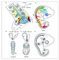
 Amphioxus Origin of the mesoderm.jpg 1.802 × 756; 793 KB
-

-

-
-
-

-
 Aves Diagrammatic transverse section of a chick embryo.jpg 1.057 × 862; 471 KB
-
 Aves FGF signalling in mesoderm migration.jpg 2.152 × 612; 371 KB
-
 Aves gastrulation Tissue flows driving primitive streak formation.jpg 1.663 × 1.621; 710 KB
-
 Aves Neural tube and somites in chick embryo.jpg 1.472 × 1.144; 674 KB
-
-

-
-
-
-

-

-

-
-
Axon-fasciculation-in-the-developing-olfactory-nerve-1749-8104-5-20-S10.ogv 20 s, 536 × 528; 7,71 MB
-
Axon-fasciculation-in-the-developing-olfactory-nerve-1749-8104-5-20-S2.ogv 27 s, 612 × 528; 10,46 MB
-
Axon-fasciculation-in-the-developing-olfactory-nerve-1749-8104-5-20-S5.ogv 20 s, 536 × 528; 8,37 MB
-
Axon-fasciculation-in-the-developing-olfactory-nerve-1749-8104-5-20-S6.ogv 18 s, 624 × 528; 9,46 MB
-
Axon-fasciculation-in-the-developing-olfactory-nerve-1749-8104-5-20-S7.ogv 20 s, 536 × 528; 9,24 MB
-
Axon-fasciculation-in-the-developing-olfactory-nerve-1749-8104-5-20-S8.ogv 17 s, 624 × 528; 6,07 MB
-
Axon-fasciculation-in-the-developing-olfactory-nerve-1749-8104-5-20-S9.ogv 17 s, 624 × 528; 7,2 MB
-
 Blastome The fate of the mesoderm and the formation of mesenchyme.jpg 1.085 × 489; 353 KB
-
 Branchiostoma formation of the neural tube, notochord, mesoderm, and coelom.jpg 1.172 × 857; 562 KB
-
-
 Caudata process of invagination and mesoderm-formation.jpg 805 × 823; 698 KB
-

-

-

-
-

-
 Characterization of the SOX2T-positive territory of the epiblast in chicken embryo.jpg 2.113 × 1.581; 1,21 MB
-
 Chordamesoderm Early processes are not affected by U0126 with the exception of apoptosis.jpg 2.146 × 3.365; 2,73 MB
-
 Coelomate 01.png 250 × 250; 12 KB
-

-
-
Control-of-Directed-Cell-Migration-In-Vivo-by-Membrane-to-Cortex-Attachment-pbio.1000544.s009.ogv 6,2 s, 264 × 264; 701 KB
-
Control-of-Directed-Cell-Migration-In-Vivo-by-Membrane-to-Cortex-Attachment-pbio.1000544.s010.ogv 6,2 s, 264 × 264; 388 KB
-
Control-of-Directed-Cell-Migration-In-Vivo-by-Membrane-to-Cortex-Attachment-pbio.1000544.s011.ogv 6,2 s, 264 × 264; 454 KB
-
Control-of-Directed-Cell-Migration-In-Vivo-by-Membrane-to-Cortex-Attachment-pbio.1000544.s012.ogv 7,6 s, 418 × 416; 1,51 MB
-
Control-of-Directed-Cell-Migration-In-Vivo-by-Membrane-to-Cortex-Attachment-pbio.1000544.s013.ogv 7,5 s, 419 × 416; 680 KB
-
Control-of-Directed-Cell-Migration-In-Vivo-by-Membrane-to-Cortex-Attachment-pbio.1000544.s014.ogv 6,5 s, 512 × 512; 532 KB
-
-
Control-of-Directed-Cell-Migration-In-Vivo-by-Membrane-to-Cortex-Attachment-pbio.1000544.s016.ogv 11 s, 1.024 × 529; 2,41 MB
-
Control-of-Directed-Cell-Migration-In-Vivo-by-Membrane-to-Cortex-Attachment-pbio.1000544.s017.ogv 5,7 s, 370 × 383; 589 KB
-
Control-of-Directed-Cell-Migration-In-Vivo-by-Membrane-to-Cortex-Attachment-pbio.1000544.s018.ogv 6,5 s, 370 × 370; 738 KB
-
-

-

-

-
 Differentiation of the mesoderm in holoblastic and meroblastic types of development.jpg 1.617 × 1.467; 1.022 KB
-
 Diversity of vertebrate gastrulation.jpg 1.073 × 1.021; 415 KB
-
 Dose-dependent reshaping of primitive streak.jpg 968 × 1.236; 743 KB
-
-
-
-
-
-
Dynamics of mesodermal cell ingression chicken embryo.ogv 12 s, 226 × 720; 4,2 MB
-
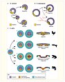
 Early gastrulation in amphibian embryos.png 3.570 × 1.358; 1.007 KB
-
Efficient-Differentiation-of-Embryonic-Stem-Cells-into-Mesodermal-Precursors-by-BMP-Retinoic-Acid-pone.0036405.s001.ogv 3 min 19 s, 480 × 360; 49,48 MB
-
 Embryologie von Physa fontinalis L. (1905) (20662318504).jpg 3.466 × 2.452; 1,44 MB
-
 Embryologie von Physa fontinalis L. (1905) (20663869963).jpg 3.544 × 1.380; 546 KB
-
 Embryologie von Physa fontinalis L. (1905) (20663883103).jpg 1.838 × 3.060; 747 KB
-
 Embryologie von Physa fontinalis L. (1905) (20663888563).jpg 2.172 × 3.170; 1.009 KB
-
 Embryologie von Physa fontinalis L. (1905) (20663916513).jpg 2.172 × 3.210; 988 KB
-
 Embryologie von Physa fontinalis L. (1905) (20663935813).jpg 3.544 × 2.566; 1,44 MB
-
 Embryologie von Physa fontinalis L. (1905) (21096895540).jpg 1.680 × 3.026; 723 KB
-
 Embryologie von Physa fontinalis L. (1905) (21096929580).jpg 3.516 × 2.544; 1,5 MB
-
 Embryologie von Physa fontinalis L. (1905) (21096946320).jpg 3.474 × 2.514; 1,59 MB
-
 Embryologie von Physa fontinalis L. (1905) (21097115988).jpg 3.412 × 2.576; 1,08 MB
-
 Embryologie von Physa fontinalis L. (1905) (21097136908).jpg 2.080 × 3.024; 1,24 MB
-
 Embryologie von Physa fontinalis L. (1905) (21097142248).jpg 2.100 × 2.954; 1,17 MB
-
 Embryologie von Physa fontinalis L. (1905) (21098108329).jpg 3.520 × 1.574; 482 KB
-
 Embryologie von Physa fontinalis L. (1905) (21098144009).jpg 3.468 × 2.434; 1,45 MB
-
 Embryologie von Physa fontinalis L. (1905) (21258748426).jpg 2.208 × 3.435; 1,08 MB
-
 Embryologie von Physa fontinalis L. (1905) (21258750436).jpg 2.166 × 2.892; 1,15 MB
-
 Embryologie von Physa fontinalis L. (1905) (21258759966).jpg 2.180 × 3.048; 1,01 MB
-
 Embryologie von Physa fontinalis L. (1905) (21258778116).jpg 2.108 × 3.004; 1,12 MB
-
 Embryologie von Physa fontinalis L. (1905) (21274404152).jpg 1.810 × 3.643; 1.012 KB
-
 Embryologie von Physa fontinalis L. (1905) (21274437582).jpg 2.068 × 2.876; 1,24 MB
-
 Embryologie von Physa fontinalis L. (1905) (21274456262).jpg 3.514 × 2.544; 1,63 MB
-
 Embryologie von Physa fontinalis L. (1905) (21285006125).jpg 2.154 × 3.074; 1,21 MB
-
 Embryologie von Physa fontinalis L. (1905) (21285013045).jpg 3.450 × 2.668; 1,44 MB
-
 Embryologie von Physa fontinalis L. (1905) (21293118491).jpg 2.182 × 3.174; 1,19 MB
-
 Embryologie von Physa fontinalis L. (1905) (21293127841).jpg 2.170 × 2.896; 1,01 MB
-
 Embryologie von Physa fontinalis L. (1905) (21293131931).jpg 3.380 × 2.300; 1,41 MB
-
 Embryologie von Physa fontinalis L. (1905) (21293139221).jpg 2.196 × 3.074; 1,07 MB
-
 Embryologie von Physa fontinalis L. (1905) (21293146721).jpg 2.080 × 3.046; 1,16 MB
-
 Embryology (1949) (21285693065).jpg 1.035 × 1.115; 581 KB
-
 Embryonic myogenesis mouse.jpg 1.261 × 1.278; 938 KB
-
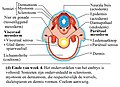 End of week 4 Embryo with somites nltxt.jpg 925 × 674; 203 KB
-
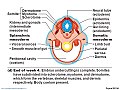 End of week 4 Embryo with somites.jpg 960 × 720; 121 KB
-
 Experimental manipulation of the gastrulation mode in different organisms.jpg 1.166 × 1.480; 850 KB
-

-

-
 Expression patterns of L. fluviatilis NogginA (a–j), NogginC (k–r) and NogginD (s–v).jpg 1.495 × 2.037; 1,69 MB
-

-
Fli+-etsrp+-Hemato-Vascular-Progenitor-Cells-Proliferate-at-the-Lateral-Plate-Mesoderm-during-pone.0014732.s008.ogv 7,2 s, 1.388 × 1.040; 1,36 MB
-
Fli+-etsrp+-Hemato-Vascular-Progenitor-Cells-Proliferate-at-the-Lateral-Plate-Mesoderm-during-pone.0014732.s009.ogv 19 s, 1.388 × 1.040; 1,13 MB
-
 Flowchart of paraxial mesodermal development and sclerotome specification.jpg 1.385 × 1.417; 673 KB
-
-
 Formation of the primitive body plan following gastrulation in the mouse.png 1.279 × 1.187; 1.016 KB
-
 Four diagrams showing hypothetical stages of early human embryos.jpg 1.631 × 1.434; 943 KB
-
From-Dynamic-Expression-Patterns-to-Boundary-Formation-in-the-Presomitic-Mesoderm-pcbi.1002586.s013.ogv 47 s, 1.272 × 322; 4,94 MB
-
From-Dynamic-Expression-Patterns-to-Boundary-Formation-in-the-Presomitic-Mesoderm-pcbi.1002586.s014.ogv 47 s, 1.272 × 322; 5,34 MB
-
From-Dynamic-Expression-Patterns-to-Boundary-Formation-in-the-Presomitic-Mesoderm-pcbi.1002586.s015.ogv 44 s, 1.272 × 322; 5,64 MB
-
From-Dynamic-Expression-Patterns-to-Boundary-Formation-in-the-Presomitic-Mesoderm-pcbi.1002586.s016.ogv 52 s, 1.272 × 322; 5,66 MB
-
From-Dynamic-Expression-Patterns-to-Boundary-Formation-in-the-Presomitic-Mesoderm-pcbi.1002586.s017.ogv 47 s, 1.272 × 322; 5,34 MB
-
From-Dynamic-Expression-Patterns-to-Boundary-Formation-in-the-Presomitic-Mesoderm-pcbi.1002586.s018.ogv 42 s, 1.272 × 322; 4,44 MB
-
From-Dynamic-Expression-Patterns-to-Boundary-Formation-in-the-Presomitic-Mesoderm-pcbi.1002586.s019.ogv 23 s, 1.277 × 274; 2,45 MB
-
From-Dynamic-Expression-Patterns-to-Boundary-Formation-in-the-Presomitic-Mesoderm-pcbi.1002586.s020.ogv 44 s, 1.277 × 160; 5,22 MB
-
From-Dynamic-Expression-Patterns-to-Boundary-Formation-in-the-Presomitic-Mesoderm-pcbi.1002586.s021.ogv 1 min 16 s, 1.272 × 322; 13,51 MB
-
From-Dynamic-Expression-Patterns-to-Boundary-Formation-in-the-Presomitic-Mesoderm-pcbi.1002586.s022.ogv 56 s, 1.272 × 322; 7,98 MB
-
 Fundulus heteroclitus presumptive organ-forming areas of the blastoderm.jpg 1.020 × 796; 779 KB
-

-

-
 Gray29.png 500 × 306; 15 KB
-

-
 Human embryo Section of embryonic rudiment in Peters' ovum (first week).jpg 1.141 × 857; 540 KB
-

-
 Human embryo Transverse Section.jpg 932 × 792; 477 KB
-

-

-
In-Silico-Patterning-of-Vascular-Mesenchymal-Cells-in-Three-Dimensions-pone.0020182.s001.ogv 15 s, 550 × 633; 1,46 MB
-
In-Silico-Patterning-of-Vascular-Mesenchymal-Cells-in-Three-Dimensions-pone.0020182.s002.ogv 15 s, 550 × 633; 1,89 MB
-
In-Silico-Patterning-of-Vascular-Mesenchymal-Cells-in-Three-Dimensions-pone.0020182.s003.ogv 14 s, 550 × 631; 669 KB
-
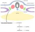 Intermediate mesoderm.png 486 × 447; 58 KB
-
 Kirkes' handbook of physiology (1907) (14769687492).jpg 1.091 × 918; 435 KB
-

-
 Location of the intermediate mesoderm nephrogenic cord..jpg 600 × 227; 58 KB
-
Long-term tracking of a nuclear red stained chicken embryo.ogv 10 s, 248 × 718; 4,63 MB
-
Long-term tracking of the mesodermal progenitors chicken embryo.ogv 10 s, 236 × 432; 889 KB
-
Longevity of tracks along the primitive streak (PS) chicken embryo.ogv 5,9 s, 286 × 720; 1,48 MB
-

-
-
-

-
Matrix-Bound-PAI-1-Supports-Cell-Blebbing-via-RhoAROCK1-Signaling-pone.0032204.s006.ogv 10 s, 309 × 320; 436 KB
-
Matrix-Bound-PAI-1-Supports-Cell-Blebbing-via-RhoAROCK1-Signaling-pone.0032204.s007.ogv 6,0 s, 240 × 240; 17 KB
-
Matrix-Bound-PAI-1-Supports-Cell-Blebbing-via-RhoAROCK1-Signaling-pone.0032204.s008.ogv 14 s, 312 × 312; 63 KB
-
Matrix-Bound-PAI-1-Supports-Cell-Blebbing-via-RhoAROCK1-Signaling-pone.0032204.s009.ogv 13 s, 240 × 240; 39 KB
-
Matrix-Bound-PAI-1-Supports-Cell-Blebbing-via-RhoAROCK1-Signaling-pone.0032204.s010.ogv 1 min 7 s, 318 × 320; 2,44 MB
-
Matrix-Bound-PAI-1-Supports-Cell-Blebbing-via-RhoAROCK1-Signaling-pone.0032204.s011.ogv 16 s, 420 × 312; 281 KB
-
Matrix-Bound-PAI-1-Supports-Cell-Blebbing-via-RhoAROCK1-Signaling-pone.0032204.s012.ogv 22 s, 418 × 320; 161 KB
-
Matrix-Bound-PAI-1-Supports-Cell-Blebbing-via-RhoAROCK1-Signaling-pone.0032204.s013.ogv 16 s, 448 × 320; 302 KB
-
 Mesoderm development.svg 512 × 197; 27 KB
-
 Mesoderm-ring to -crescent transition.jpg 1.173 × 1.826; 1,01 MB
-
 Neurula.png 873 × 317; 21 KB
-
 Not all tissues receiving notochord-emitted signals are affected by U0126 treatment.jpg 2.362 × 1.496; 1,48 MB
-
 Overview of iPS cells.png 478 × 361; 13 KB
-
 Overview of the mesodermal derivatives.jpg 1.494 × 1.147; 656 KB
-
 Pedicellina echinata Early stages in the development of the egg.jpg 1.278 × 844; 239 KB
-

-
 Primitiv Node.jpg 720 × 504; 42 KB
-
 Primitive Trilaminar Human Embryo in Tubal Pregnancy (40X) (3944578509).jpg 1.712 × 1.206; 875 KB
-
-
-
-
-
-
 Salmo irideus presumptive organ-forming areas in the blastoderm.jpg 1.163 × 884; 685 KB
-

-

-
-

-

-
-

-
Specific-Syndecan-1-Domains-Regulate-Mesenchymal-Tumor-Cell-Adhesion-Motility-and-Migration-pone.0014816.s002.ogv 8,1 s, 1.384 × 1.038; 6,41 MB
-
Specific-Syndecan-1-Domains-Regulate-Mesenchymal-Tumor-Cell-Adhesion-Motility-and-Migration-pone.0014816.s003.ogv 8,1 s, 1.384 × 1.038; 3,64 MB
-
Specific-Syndecan-1-Domains-Regulate-Mesenchymal-Tumor-Cell-Adhesion-Motility-and-Migration-pone.0014816.s004.ogv 8,1 s, 1.384 × 1.038; 2,87 MB
-
Specific-Syndecan-1-Domains-Regulate-Mesenchymal-Tumor-Cell-Adhesion-Motility-and-Migration-pone.0014816.s005.ogv 8,1 s, 1.384 × 1.038; 3,18 MB
-
Specific-Syndecan-1-Domains-Regulate-Mesenchymal-Tumor-Cell-Adhesion-Motility-and-Migration-pone.0014816.s006.ogv 8,1 s, 1.384 × 1.038; 4,2 MB
-
Specific-Syndecan-1-Domains-Regulate-Mesenchymal-Tumor-Cell-Adhesion-Motility-and-Migration-pone.0014816.s007.ogv 8,0 s, 1.024 × 1.024; 2,83 MB
-
Specific-Syndecan-1-Domains-Regulate-Mesenchymal-Tumor-Cell-Adhesion-Motility-and-Migration-pone.0014816.s008.ogv 8,0 s, 1.024 × 1.024; 2,36 MB
-
Specific-Syndecan-1-Domains-Regulate-Mesenchymal-Tumor-Cell-Adhesion-Motility-and-Migration-pone.0014816.s009.ogv 8,0 s, 1.024 × 1.024; 1,73 MB
-
Specific-Syndecan-1-Domains-Regulate-Mesenchymal-Tumor-Cell-Adhesion-Motility-and-Migration-pone.0014816.s010.ogv 8,0 s, 1.024 × 1.024; 5,97 MB
-
Specific-Syndecan-1-Domains-Regulate-Mesenchymal-Tumor-Cell-Adhesion-Motility-and-Migration-pone.0014816.s011.ogv 8,0 s, 1.024 × 1.024; 3,46 MB
-
-


_epiblast_chicken_embryo.jpg)


_(20047812224).jpg)
_(20661197932).jpg)


,_myotome_(muscle-producer),_and_sclerotome_(skeleton-producer).jpg)


_epithelial_phenotype_during_development_chicken_embryo.jpg)
_(20662318504).jpg)
_(20663883103).jpg)
_(20663888563).jpg)
_(20663916513).jpg)
_(20663935813).jpg)
_(21096895540).jpg)
_(21096929580).jpg)
_(21096946320).jpg)
_(21097115988).jpg)
_(21097136908).jpg)
_(21097142248).jpg)
_(21098108329).jpg)
_(21098144009).jpg)
_(21258748426).jpg)
_(21258750436).jpg)
_(21258759966).jpg)
_(21258778116).jpg)
_(21274404152).jpg)
_(21274437582).jpg)
_(21274456262).jpg)
_(21285006125).jpg)
_(21285013045).jpg)
_(21293118491).jpg)
_(21293127841).jpg)
_(21293131931).jpg)
_(21293139221).jpg)
_(21293146721).jpg)
_(21285693065).jpg)


_signature_genes_in_chicken_and_mouse_embryos.jpg)
.jpg)
,_NogginC_(k%E2%80%93r)_and_NogginD_(s%E2%80%93v).jpg)


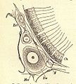
-_section_o,_a,_calcareous_sponge_Ect_ectoderm;_Mes_mesoderm;_N,_calcerous_spicule;_Eis.jpeg)


.jpg)


_(14769687492).jpg)


_progenitor_territories_results_in_NMPs_remaining_as_the_major_PS_remnant_in_the_tail_bud.jpg)

_(3944578509).jpg)


_analysis_of_the_posterior_tissue_precursors_during_posterior_axis_formation_chicken_embryo.jpg)
_marker_tbx20_during_zebrafish_early_development.jpg)
_during_zebrafish_early_development.jpg)
_during_zebrafish_early_development.jpg)
_during_zebrafish_early_development.jpg)
_(20663869963).jpg)