Category:Microtomes
Salti al navigilo
Salti al serĉilo
system to cut fine samples for microscopy  | |||||
| Alŝuti plurmedion | |||||
| Subaro de | |||||
|---|---|---|---|---|---|
| Uzado | |||||
| |||||
Subkategorioj
Ĉi tiu kategorio havas la 5 jenajn subkategoriojn, el 5 entute.
C
- Cryotomy (15 D)
H
M
- Microtome (Literature) (9 D)
- Microtome-MHS 428 (6 D)
U
- Ultramicrotomes (21 D)
Dosieroj en kategorio “Microtomes”
La jenaj 61 dosieroj estas en ĉi tiu kategorio, el 61 entute.
-
 A freezing microtome of 1873. Wellcome M0016984.jpg 5 220 × 2 023; 1,7 MB
-
 Base sledge microtome.jpg 640 × 393; 45 KB
-
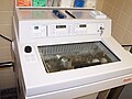
 Blick in Kryostat für histologische Schnitte.jpg 5 184 × 3 888; 4,53 MB
-

-
 Cryostat microtome.jpg 800 × 600; 67 KB
-
 CSIRO ScienceImage 1915 The Immunohistochemistry Laboratory.jpg 1 772 × 1 408; 1,55 MB
-
 Cummings 1774 Microtome.jpg 1 434 × 2 772; 1,81 MB
-
 Cutting station.jpg 1 536 × 2 304; 552 KB
-
 EB1911 Microtomy - Large Sliding Microtome.jpg 963 × 701; 256 KB
-
 Electrical microtome.jpg 1 548 × 1 916; 1 000 KB
-

-
-
 Freezing microtome, London, England, 1883-1885 Wellcome L0058209.jpg 4 256 × 2 832; 1,25 MB
-
 Hand Microtome.png 751 × 475; 604 KB
-
 His 1870 Mikrotom (Bild 1).png 2 608 × 3 576; 2,35 MB
-
 His 1870 Mikrotom (Bild 2).png 2 777 × 1 799; 592 KB
-
 His 1870 Mikrotom.png 2 362 × 5 033; 2,45 MB
-
 Home-made-microtome.svg 1 213 × 2 038; 38 KB
-
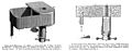
 Journal of applied microscopy and laboratory methods (1901) (14597383809).jpg 2 350 × 3 613; 907 KB
-
 Kryostat für histologische Schnitte.jpg 5 184 × 3 888; 4,54 MB
-
 Laser-microtome-schematic.png 785 × 501; 74 KB
-
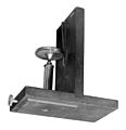
 Leake microtome.jpg 680 × 462; 89 KB
-
 Leica microtome 2.JPG 2 448 × 3 264; 2,2 MB
-
 Leica microtome.JPG 2 448 × 3 264; 1,92 MB
-
 Leica SM2000R-Microtome.jpg 3 840 × 5 120; 3,14 MB
-
 Messerstellung (Deklination) bei der Mikrotomie.svg 540 × 760; 14 KB
-
 Messerstellung (Inklination) bei der Mikrotomie.svg 600 × 360; 16 KB
-
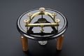
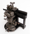
 Microtome - Cabinet of Physics (Arppeanum) - - DSC05170.JPG 4 320 × 3 240; 4,53 MB
-
 Microtome de Minot.jpg 4 803 × 3 841; 5,45 MB
-
 Microtome of the type used by Quekett. Wellcome M0010835.jpg 3 578 × 3 108; 2,45 MB
-
 Microtome principle.svg 550 × 500; 22 KB
-
 Microtome used in the 1850's. Wellcome M0018210.jpg 3 201 × 3 298; 889 KB
-
 Microtome-1.jpg 1 146 × 1 284; 676 KB
-
 Microtome-knife-profile.svg 350 × 600; 5 KB
-
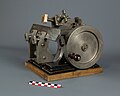
 Microtome1905.JPG 3 696 × 2 247; 3,56 MB
-
 Microtomo a slitta.jpg 3 264 × 2 448; 754 KB
-
 Microtomo rotativo.jpg 1 248 × 1 296; 188 KB
-
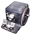
 Microtomo.jpg 1 240 × 1 240; 92 KB
-
 Mikrotom 02.jpg 2 048 × 1 536; 1,67 MB
-
 Mikrotom R-Jung Heidelberg.png 595 × 681; 340 KB
-
 Mikrotom-sartorius hg.jpg 2 417 × 2 675; 627 KB
-
 Mikrotom-Schlifftypen.svg 350 × 600; 6 KB
-
 Mikrotom.jpg 2 752 × 2 832; 584 KB
-
 Mikrotom01.jpg 2 048 × 1 536; 1,92 MB
-
 OSC Microbio 02 04 Microtome.jpg 900 × 369; 191 KB
-
 Prinzip Lasermikrotom.jpg 785 × 501; 81 KB
-
 Prinzip Mikrotomschnitt (Rotationsmikrotom).svg 550 × 500; 24 KB
-
 Rocking microtome, Cambridge, England Wellcome L0059119.jpg 4 256 × 2 832; 1,45 MB
-
 Rotary microtome in action.jpg 576 × 436; 40 KB
-

-
 Sledemicrotoom Reichert-Jung Hn40.jpg 1 957 × 1 724; 686 KB
-
 Sledge microtome.jpg 576 × 436; 46 KB
-

-
 The diagnosis of diseases of women (1905) (14760034431).jpg 1 752 × 1 938; 571 KB
-
 The diagnosis of diseases of women (1905) (14762881582).jpg 2 128 × 1 390; 583 KB
-
 The Microtome, designed by Wilhem His. Wellcome M0010853.jpg 2 780 × 4 010; 1,92 MB
-
 Tissue chopper.jpg 5 184 × 3 888; 2,69 MB
-

-
 Tissue processing - Solid paraffin block is mounted on the tissue holder of a microtome.jpg 4 288 × 2 848; 4,71 MB
-
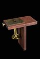 Topping-type microtome, Europe, 1840-1850 Wellcome L0058210.jpg 2 676 × 3 921; 1,21 MB
-
 Одноразовое лезвие для микротома.jpg 5 448 × 3 632; 9 MB


.jpg)


.png)
.png)


_(14597383809).jpg)


_bei_der_Mikrotomie.svg)
_bei_der_Mikrotomie.svg)
_-_-_DSC05170.JPG)


.svg)


_(14760034431).jpg)
_(14762881582).jpg)


