Category:Mycelium
Salti al navigilo
Salti al serĉilo
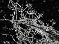
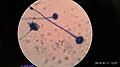
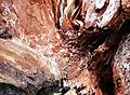
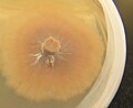
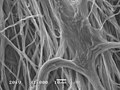
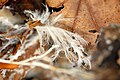
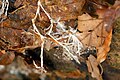
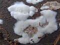
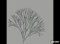
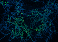
Mycelium is the vegetative part of a fungus, consisting of a mass of branching, thread-like hyphae.
Internationalization
Deutsch: Myzel
· Español: Micelio
· Français : Mycélium
· Italiano: Micelio
· Nederlands: Zwamvlok
· Română: Miceliu
· Русский: Мицелий
· the vegetative part of a fungus, consisting of a mass of branching, thread-like hyphae  | |||||
| Alŝuti plurmedion | |||||
| Estas |
| ||||
|---|---|---|---|---|---|
| Parto de | |||||
| |||||
Subkategorioj
Ĉi tiu kategorio havas la 2 jenajn subkategoriojn, el 2 entute.
H
M
- Mycelia on photographic lenses (13 D)
Dosieroj en kategorio “Mycelium”
La jenaj 94 dosieroj estas en ĉi tiu kategorio, el 94 entute.
-
 A common mycelium.jpg 3 096 × 4 128; 2,03 MB
-
 A-sexy mycelium.jpg 1 684 × 1 268; 124 KB
-
 Annual report of the State Botanist of the State of New York (1887) (19179744580).jpg 1 700 × 2 836; 1,72 MB
-

-
 Armillaria spp Mycelia Mats.jpg 768 × 511; 327 KB
-
 Ashby Plot.png 998 × 386; 204 KB
-
 Atti BHL3917595.jpg 2 021 × 3 105; 403 KB
-
 Beneath, by Beth Walker (UK) at 2022 Fungi Film Festival.jpg 2 400 × 1 350; 322 KB
-

-
 Bridge of cells.jpg 2 048 × 1 536; 636 KB
-
 Competitive contamination.jpg 2 048 × 1 536; 757 KB
-
-
 Coniophora puteana - Lindsey.jpg 640 × 426; 83 KB
-
 D'une spore au mycélium.jpg 1 389 × 328; 68 KB
-
De-Myzel.ogg 2,0 s; 19 KB
-
De-Myzelium.ogg 1,8 s; 18 KB
-
 Department bulletin (1900) (20671560820).jpg 2 416 × 3 924; 1,25 MB
-
 Diagram of mycelium based acoustic panel production.jpg 1 843 × 813; 1,01 MB
-
 DSC0727 Wolfgang Günzel.jpg 4 724 × 3 218; 15,3 MB
-
 Elementary botany (1905) (14596667099).jpg 2 860 × 1 536; 1,01 MB
-

-

-
 Figure 5 in 'Mind the Fungi' by Meyer & Pfeiffer (2) - picture by Wolfgang Günzel.jpg 3 632 × 5 319; 14,91 MB
-
 Figure 5 in 'Mind the Fungi' by Meyer & Pfeiffer (3) - picture by Wolfgang Günzel.jpg 2 560 × 3 840; 3,1 MB
-
 Figure 5 in 'Mind the Fungi' by Meyer & Pfeiffer - picture by Wolfgang Günzel.jpg 5 760 × 3 840; 15,23 MB
-

-
 Foreign Exchange (Charon) .jpg 2 448 × 3 264; 1,53 MB
-
 Formacion de sinemas.jpg 1 333 × 749; 113 KB
-
 Gaillard's medical journal (1882) (14585726379).jpg 2 166 × 3 238; 574 KB
-
 Grubenholz Bewuchs Philippstollen 2.jpg 4 000 × 3 000; 1,44 MB
-
 Grubenholz Bewuchs Philippstollen 3.jpg 3 000 × 4 000; 1,47 MB
-
 Grubenholz Bewuchs Philippstollen.jpg 4 000 × 3 000; 1,56 MB
-
 Grzybnia - mycelium.jpg 2 736 × 2 052; 746 KB
-
 Grzybnia immobilizowana.jpg 4 608 × 3 456; 3,84 MB
-
 Grzybnia żółciaka siarkowego w śliwie.jpg 4 336 × 3 168; 3,41 MB
-
 Heksenring.jpg 4 000 × 3 000; 1,71 MB
-
 Lactarius deliciosus in Petri.JPG 1 769 × 1 450; 850 KB
-
 Laetiporus sulphureus mycelium.jpg 4 140 × 3 188; 3,19 MB
-
 Micelio en forma circular en PDA.jpg 1 285 × 722; 117 KB
-
 Micelio y carpóforos de champiñón (cropped).JPG 2 628 × 1 848; 1,86 MB
-
 Micelio y carpóforos de champiñón en un cultivo de Pradejón.jpg 3 888 × 2 592; 6,81 MB
-
 Micelio y carpóforos de champiñón.JPG 2 704 × 4 064; 3,55 MB
-
 Mushroom's roots (mycélium).jpg 800 × 600; 105 KB
-

-
 Mycelia of Piptocephalis Freseniana.jpg 1 130 × 1 711; 413 KB
-
 Mycelial cord Armillaria (rhizomorphs).jpg 5 152 × 3 864; 5,78 MB
-
 Mycelial film.jpg 1 280 × 960; 449 KB
-
 Mycelium (4).jpg 3 968 × 2 976; 2,77 MB
-
 Mycelium (5).jpg 3 968 × 2 976; 2,7 MB
-
 Mycelium (6).jpg 3 968 × 2 976; 2,72 MB
-
 Mycelium - Lindsey 1a.jpg 640 × 428; 118 KB
-
 Mycelium - Lindsey 1b.jpg 640 × 428; 154 KB
-
 Mycelium a1(2).jpg 1 950 × 1 462; 776 KB
-
 Mycelium growth simulation.gif 492 × 360; 2,22 MB
-
 Mycelium growth, Chapeltoun, North Ayrshire.jpg 2 898 × 4 134; 2,52 MB
-
 Mycelium in forest floor.jpg 1 280 × 1 266; 676 KB
-
 Mycelium objects at Genspace (75903).jpg 4 244 × 2 271; 7,56 MB
-
 Mycelium objects at Genspace (75904).jpg 2 325 × 2 449; 5,57 MB
-
 Mycelium objects at Genspace (75906).jpg 4 553 × 3 318; 11,18 MB
-
 Mycelium of arbuscular mycorrhizal fungi with false color.png 700 × 513; 553 KB
-
-
 Mycelium Pleurotus citrinopileatus R.H..jpg 3 968 × 2 976; 2,73 MB
-
 Mycelium ramifying over stick under beech leaves. Garth 1971 (22843228098).jpg 1 500 × 974; 237 KB
-
 Mycelium RH (1).jpg 3 968 × 2 976; 2,75 MB
-
 Mycelium RH (3).jpg 3 968 × 2 976; 2,78 MB
-
 Mycelium RH (6).jpg 3 968 × 2 976; 2,8 MB
-
 Mycelium RH (8).jpg 3 968 × 2 976; 2,74 MB
-
 Mycelium RH (9).jpg 3 968 × 2 976; 2,82 MB
-
 Mycelium-001 (8260778720).jpg 1 600 × 1 067; 752 KB
-
 Mycélium au devant de la scène.jpg 1 584 × 1 056; 905 KB
-

-
 Oyster mushroom (Pleurotus ostreatus) mycelium on coffee grounds.JPG 1 024 × 768; 255 KB
-
 Panellus Stipticus, Bioluminescent Fungi Spores 01.jpg 2 602 × 3 908; 1,76 MB
-
 Panellus Stipticus, Bioluminescent Fungi Spores 02.jpg 3 908 × 2 602; 2,47 MB
-
 Panellus Stipticus, Bioluminescent Fungi Spores 03.jpg 3 908 × 2 602; 4,28 MB
-
 Parental Advice.jpg 2 048 × 1 536; 740 KB
-
 Phillipsstollen, Mycel auf Wand und Holz, Sauerland GLAM 359 3 PK.jpg 2 417 × 2 596; 1,89 MB
-
 Phillipsstollen, Pilzmyzel, Sauerland GLAM 359 1 PK.jpg 7 360 × 4 912; 10,04 MB
-
 Pleurotus ostreatus mycelium on coffee grounds.JPG 463 × 317; 92 KB
-
 Pleurotus ostreatus on agar.jpg 2 612 × 2 918; 1,63 MB
-
 Pleurotus ostreatus R.H.1.jpg 3 968 × 2 976; 2,76 MB
-
 Reproduction sexuée des basidiomycètes.jpg 855 × 630; 79 KB
-
 Rhizomorph Armillaria 2PN.jpg 2 792 × 2 094; 2,19 MB
-
 Rhizoplanen Pilze.tif 4 398 × 2 831; 1 001 KB
-
 Sporangia Myceliun.jpg 1 024 × 768; 307 KB
-
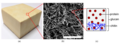
 Stress Strain curves (a) uniaxial tension and (b) compression.png 841 × 354; 97 KB
-
 T. versicolor- mycelium.jpg 1 920 × 2 560; 2,27 MB
-
 The root-like Mycelium of a fungus.jpg 2 268 × 4 032; 2,18 MB
-

-
 White mycelium growing on agar.jpg 2 448 × 3 264; 1,19 MB
-
 X hypoxylon.jpg 1 536 × 2 023; 327 KB
-
 Śliwa po uderzeniu pioruna z żółciakiem.jpg 2 792 × 2 944; 2,08 MB
-
 Гриб рода Mucor под электронным микроскопом.jpg 1 836 × 2 075; 741 KB
-
 Мицелија на габата Fusarium sp.jpg 720 × 576; 403 KB


_(19179744580).jpg)


_at_2022_Fungi_Film_Festival.jpg)


_(20671560820).jpg)


_(14596667099).jpg)

.jpg)
_-_picture_by_Wolfgang_G%C3%BCnzel.jpg)
_-_picture_by_Wolfgang_G%C3%BCnzel.jpg)

_.jpg)

_(14585726379).jpg)


.JPG)


.jpg)


.jpg)

.jpg)
.jpg)
.jpg)


.jpg)


.jpg)
.jpg)
.jpg)


.jpg)
.jpg)
.jpg)
.jpg)
.jpg)
.jpg)
.jpg)

_mycelium_in_petri_dish_on_coffee_grounds.JPG)
_mycelium_on_coffee_grounds.JPG)


_uniaxial_tension_and_(b)_compression.png)


_and_is_laying_down_after_the_storm_at_Schaarsbergen_-_panoramio.jpg)


_Current_state_and_future_prospects_of_pure_mycelium_materials.jpg)
_macroscale,_(b)_SEM_image_with_100_micron_scale_bar,_(c)_microstructure.png)