Category:Neuroanatomy
Zur Navigation springen
Zur Suche springen
English: Neuroanatomy is the branch of anatomy that studies the anatomical organization of the nervous system.
Teilgebiet der Anatomie, das sich mit dem Nervensystem beschäftigt | |||||
| Medium hochladen | |||||
| Ist ein(e) | |||||
|---|---|---|---|---|---|
| Ist Teil von | |||||
| |||||
Unterkategorien
Es werden 20 von insgesamt 20 Unterkategorien in dieser Kategorie angezeigt:
In Klammern die Anzahl der enthaltenen Kategorien (K), Seiten (S), Dateien (D)
Medien in der Kategorie „Neuroanatomy“
Folgende 117 Dateien sind in dieser Kategorie, von 117 insgesamt.
-
-
 1409 Maculae and Equilibrium.jpg 2.388 × 1.254; 1,05 MB
-
 1410 Equilibrium and Semicircular Canals.jpg 2.256 × 1.112; 809 KB
-
 1418 Auditory Brainstem Mechanisms.jpg 1.033 × 2.560; 607 KB
-
 1504 Autonomic Varicosities.jpg 1.971 × 846; 549 KB
-
 1505 Comparison of Somatic and Visceral Reflexes.jpg 1.983 × 2.717; 1,38 MB
-
 Amigdala aurre-albotik.tif 1.048 × 603; 219 KB
-
 Amigdala Gorputza aurreko ikuspegia.tif 1.048 × 603; 162 KB
-
 An Atlas of the Medulla and Midbrain Plate III (high res).jpg 4.005 × 5.858; 18,59 MB
-
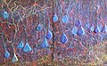
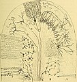
 Anatomie des centres nerveux (1895) (14788115633).jpg 1.674 × 2.090; 732 KB
-
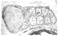
 Area postrema micrograph.jpg 2.400 × 2.254; 3,12 MB
-
 Auditory Tract.tif 2.347 × 3.510; 5,95 MB
-
Axonal nerve fibers in a brain - the neural network that is us.webm 14 s, 964 × 720; 2,99 MB
-
 Basalganglia.svg 524 × 334; 9 KB
-
 Basic neuroanatomy of the gustatory system.jpg 425 × 600; 161 KB
-
 BedNucleusST.png 942 × 822; 804 KB
-
 Beheko oliba nukleoa.tif 1.602 × 916; 244 KB
-
 Brain layers.jpg 382 × 294; 28 KB
-
 CA1 to subiculum4.jpg 1.010 × 624; 249 KB
-
 Cajal 1898 Fig6.png 1.370 × 1.524; 654 KB
-
 Capsula interna.tif 2.347 × 3.510; 5,67 MB
-
 Cavum septi pellucidi fetal.jpg 802 × 1.161; 145 KB
-
 Cerebellar Tract.tif 2.347 × 3.510; 4,48 MB
-
 Cerebral Cortex Wiring.tif 2.347 × 3.510; 3,69 MB
-
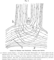
 Christian and Thompson Figure 5 SVG.svg 990 × 765; 39 KB
-
 Cognitive science heptagram tr.svg 1.578 × 1.809; 150 KB
-
 Cognitive science heptagram.svg 1.578 × 1.809; 116 KB
-
 Connectome.jpg 964 × 720; 426 KB
-
 Corticalorient.GIF 450 × 327; 5 KB
-
 Cranial Nerve Nuclei.tif 2.293 × 1.918; 2,5 MB
-
 Der Bau des centralen Nervensystemes der ungeschwänzten Batrachier.jpg 2.412 × 3.366; 631 KB
-
 Die Funktionen des Centralnervensystems und ihre phylogenese (1900) (20917206915).jpg 1.218 × 2.902; 269 KB
-
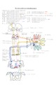
 Dor e temperatura.png 1.468 × 908; 144 KB
-
 Einstein brain.jpg 1.022 × 348; 139 KB
-
 Face antérieure du tronc cérébral.jpg 1.700 × 2.338; 1,69 MB
-
 Face latérale du tronc cérébral.jpg 1.700 × 2.338; 1,41 MB
-
 Face postérieure du tronc cérébral.jpg 1.700 × 2.338; 1,8 MB
-
 FoxP2+TH sagittal.jpg 1.973 × 1.614; 485 KB
-
 Fr-Cognitive science heptagram.svg 1.600 × 1.809; 161 KB
-
 Gehirn kompakt.png 2.956 × 1.105; 629 KB
-
 Gyri of lateral cortex.png 2.400 × 2.400; 3,11 MB
-
 Horizontal section of mibrain-superior colliculus-ja.svg 1.220 × 580; 146 KB
-
 Kasraie scalped 3d010r cal BET2 0001.png 405 × 405; 61 KB
-
 Kea0003-papezkreis.PNG 584 × 254; 21 KB
-
 Kleinhirn pontocerebellum.png 567 × 667; 51 KB
-
 Kleinhirn spinocerebellum.png 534 × 655; 48 KB
-
 Kleinhirn vestibulocerebellum.png 504 × 685; 50 KB
-
 Kortex schichten.jpg 783 × 580; 147 KB
-
 Lawrence 1960 1.3 A.png 1.136 × 1.740; 173 KB
-
 Lawrence 1960 1.4 A.png 2.160 × 750; 146 KB
-
 Lawrence 1960 21.11.png 2.396 × 2.048; 968 KB
-
 Lawrence 1960 21.16.png 2.168 × 1.908; 934 KB
-
 Lawrence 1960 6.10.png 2.252 × 1.380; 1,1 MB
-
 Lawrence 1960 6.11.png 1.120 × 572; 270 KB
-
 Lawrence 1960 6.12.png 2.248 × 1.708; 1,43 MB
-
 Lawrence 1960 6.13.png 2.144 × 2.932; 2,35 MB
-
 Lawrence 1960 6.2.png 2.416 × 1.360; 124 KB
-
 Lawrence 1960 6.3.png 2.120 × 1.352; 107 KB
-
 Lawrence 1960 6.4.png 508 × 1.164; 205 KB
-
 Lawrence 1960 6.5.png 1.072 × 776; 399 KB
-
 Lawrence 1960 6.6.png 2.172 × 1.312; 796 KB
-
 Lawrence 1960 6.7.png 2.168 × 1.272; 971 KB
-
 Lawrence 1960 6.8.png 2.188 × 2.528; 2,3 MB
-
 Lawrence 1960 6.9.png 1.116 × 1.128; 360 KB
-

-

-
-
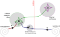 LSO wiring.png 500 × 305; 34 KB
-
 LSO wiring.svg 1.025 × 625; 108 KB
-
 MANGO histogram 0001.png 522 × 237; 2 KB
-
 MAP2-tau in neurons.jpg 926 × 935; 1,56 MB
-
 Mechanism of Ejaculation.jpg 960 × 720; 71 KB
-
 Medula espinhal cervical com raízes.png 1.801 × 432; 72 KB
-
 Medulla spinalis.tif 2.390 × 3.510; 5,09 MB
-
 Mesencephalon Section.tif 2.293 × 2.550; 3,19 MB
-
 Meynert1885.PNG 1.400 × 867; 1,1 MB
-
 Microanatomy and Wiring of Cerebellum.tif 2.261 × 3.075; 5,38 MB
-
 Moelle-Substance Blanche-Les Voies.svg 854 × 650; 58 KB
-
 Monakow atlas.JPG 614 × 1.582; 192 KB
-
 Monro, Winslow, Innes. A system of anatomy - vol 2 Wellcome L0032378.jpg 3.048 × 5.137; 6,42 MB
-
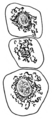
 Nerve cells. Wellcome L0001966.jpg 1.056 × 1.772; 691 KB
-
 Nervo pelo braço.png 1.724 × 1.224; 95 KB
-
 Nervous plexi.jpg 760 × 1.014; 116 KB
-
 Neuron undergoing chromatolysis.jpg 722 × 680; 83 KB
-
 Papez Circuit-pl.svg 1.440 × 1.246; 25 KB
-
 Papez Circuit.jpg 1.390 × 1.198; 157 KB
-
 Papezův okruh.svg 478 × 505; 31 KB
-
 PSM V75 D148 Differentiation of neuromuscular constituents.png 1.048 × 893; 26 KB
-
 PSM V75 D148 Neuromuscular cell.png 608 × 244; 6 KB
-
 PVH-PHAL+TH+ChAT sagittal.jpg 1.835 × 1.335; 396 KB
-
 Rhinalflow.gif 600 × 122; 17 KB
-
 Sheep midbrain.jpg 1.200 × 1.075; 334 KB
-
 StrokeMCA overlay.png 820 × 958; 340 KB
-
 Substantia nigra M mulatta.tif 900 × 581; 1,13 MB
-
 Sulcus centralis - Identification axial - MRI T2.jpg 681 × 795; 69 KB
-
 Sulcus centralis - Identification sagittal - MRI T2.jpg 912 × 700; 66 KB
-
 Temporal droit2.png 600 × 534; 129 KB
-
 Thalamus anteriore mediale laterale kerngruppe2.jpg 506 × 902; 76 KB
-
 Thalamus dorsale kerngruppe2.jpg 514 × 906; 77 KB
-
 Thalamus ventrale kerngruppe2.jpg 493 × 642; 48 KB
-

-

-

-

-

-

-

-
 The nervous system; two figures showing the brain, spine and Wellcome V0008430EL.jpg 1.254 × 2.170; 1,87 MB
-
 Tętnice mózgowe angio-MRI.jpg 663 × 362; 32 KB
-
 Vertebrate-brain-cartoon cs.png 681 × 270; 52 KB
-
 Vestibular balance system.jpg 1.568 × 948; 174 KB
-
 Vias ascendentes.png 1.352 × 752; 71 KB
-
 Vomer1.png 564 × 427; 34 KB
-
 Vue externe des noyaux gris centraux.jpg 1.700 × 2.338; 1,32 MB
-
 Vue interne des noyaux gris centraux.jpg 1.700 × 2.338; 1,74 MB
-

-
 Гіпокампна формація та медіальна енторіальна кора щура..png 1.031 × 1.371; 400 KB
.jpg)


.jpg)
_(14788115633).jpg)


_(20917206915).jpg)


_(14596358647).jpg)
_(14780531334).jpg)
_(14802722683).jpg)


_(17571976554).jpg)
_(17572193254).jpg)
_(17573537913).jpg)
_(18167660926).jpg)
_(18168333416).jpg)
_(18190879172).jpg)
_(18194518975).jpg)


