Category:Nuclear medicine
Salta a la navigazzion
Và a cercà
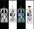
specialità medica  Un examen de médecine nucléaire « corps entier ». | |||||
| Carega su un fail audio / video | |||||
| L'è un(a) | |||||
|---|---|---|---|---|---|
| Sotoclase de |
| ||||
| |||||
This category is for images about principial of nuclear medicine. A sub-category of medicine.
See also:
Sotocategorie
Quella categoria chì la gh'ha 15 sot-categorie chì de sota, su un numer complessiv de 15.
D
- Dacryoscintigraphy (2 F)
H
M
- Media from EJNMMI Research (12 F)
- MUGA scan (2 F)
N
P
- PET/CT imaging (2 F)
R
S
Fail in la categoria "Nuclear medicine"
Quella categoria chì la gh'ha denter 125 i fail riportad chì de sota, su un total de 125.
-
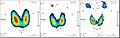
 Abnl petct.jpg 995 × 859; 87 KByte
-
 Accl2 ric.jpg 1 600 × 1 098; 261 KByte
-
 AdditionOfRepsonseFunctionsOfKidneysAndTissiueToATotalResponseFunction-de.svg 2 730 × 1 930; 208 KByte
-
 Aktivimeter Bedienoberfläche.JPG 2 492 × 1 951; 788 KByte
-
 Amyloid deposition (DPD scan).png 587 × 381; 63 KByte
-
 Amyloid deposition diagnosis 01.png 1 200 × 1 077; 1,01 MByte
-
 AnalysesOfAMAG3Renogram.png 921 × 894; 413 KByte
-
 Background corrected renal curves from a patients renogram-de.svg 608 × 558; 23 KByte
-
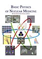
 Basic Physics of Nuclear Medicine.pdf 1 275 × 1 650, 109 pagine; 1,95 MByte
-
 Bildgebung des Gehirns und Kognition.jpg 633 × 903; 63 KByte
-
 Bleibehaelter Fluessigiod.jpg 1 370 × 1 353; 1,02 MByte
-
 BOMAB phantom2.JPG 321 × 515; 64 KByte
-
 BOMAB2.JPG 198 × 590; 10 KByte
-
 BpnmFrontCover2.jpg 562 × 793; 55 KByte
-
 Brachytherapy.jpg 4 032 × 3 024; 4,56 MByte
-
 Caméras et images de médecine nucléaire.jpg 396 × 240; 20 KByte
-
-
-
 CNX Chem 21 00 NuclearMed.png 1 300 × 600; 1,39 MByte
-
 CNX Chem 21 05 Tc-99.jpg 650 × 361; 894 KByte
-
 CNX Chem 21 05 Thallium.jpg 650 × 683; 411 KByte
-
 Curve salivare normale1.jpg 1 069 × 712; 183 KByte
-
 Diagnostics-09-00012-g011.jpg 737 × 442; 91 KByte
-
 Dinamica scintigrafia ghiandole salivari.jpg 1 075 × 727; 98 KByte
-
 DMSA scan while on enalapril.jpg 600 × 577; 357 KByte
-
 Dose Calibrator Diagram.png 2 175 × 994; 129 KByte
-
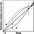 Dose-effect laws.jpg 1 790 × 1 800; 234 KByte
-
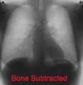 DualEnergyAbsorptiometryofChestBoneSubtracted.png 199 × 204; 31 KByte
-
 DualEnergyAbsorptiometryofChestTissueSubtracted.png 199 × 204; 45 KByte
-
 Dózisok.jpg 626 × 380; 72 KByte
-
 Espectro RMN COSY.png 763 × 726; 56 KByte
-
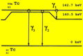
 Esquema do decaimento do Mo-99 para o Tc-99m.svg 528 × 193; 10 KByte
-
 Estehaleh Tc99m.png 588 × 396; 30 KByte
-
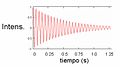
 ExponentialDecayWithTwoDifferentExponents-de.svg 820 × 580; 206 KByte
-
 FBP Iter single.jpg 968 × 975; 43 KByte
-
Feasibility-and-Safety-of-Fiber-Optic-Micro-Imaging-in-Canine-Peripheral-Airways-pone.0084829.s002.ogv 32 s, 720 × 576; 1,06 MByte
-
 Fid.jpg 614 × 345; 17 KByte
-
 Figure 1 (7001447267).png 898 × 580; 751 KByte
-
 Filtro paso de baja.png 702 × 470; 116 KByte
-
 Filtros paso de alta.png 709 × 470; 123 KByte
-
 Filtros paso de baja.png 702 × 470; 117 KByte
-
 FiveCompartmentModelInRenography-de.svg 1 200 × 2 040; 21 KByte
-
 GeneralFormOfTheImpulseResponseFunctionInDeconvolutionRenography-de.svg 1 105 × 1 005; 76 KByte
-
 Gerador de Tecnécio-99m.svg 591 × 419; 39 KByte
-
 IGAS Rapport.png 1 240 × 1 755; 855 KByte
-
 IllustrationOfTheResponseToNonIdealIInputInDeconvolutionRenography-de.svg 500 × 500; 70 KByte
-
 Lead pig2.jpg 540 × 595; 77 KByte
-
 Lead pig3.jpg 459 × 572; 59 KByte
-
 Linfoscintigrafia AAII.jpg 1 072 × 630; 199 KByte
-
 Linfoscintigrafia AASS.jpg 724 × 466; 99 KByte
-
 Logarithm of bzero.gif 786 × 619; 22 KByte
-
 LogscalePlotToCalculateRenalClearance-de-1.svg 1 102 × 673; 117 KByte
-
 LogscalePlotToCalculateRenalClearance-de.svg 1 102 × 673; 116 KByte
-
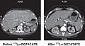 Lutetium-177 treatment CT scan.jpg 1 667 × 912; 220 KByte
-
 Mammography and scintimammography of carcinoma.jpg 1 004 × 565; 159 KByte
-
 Marinelli-NTIS Biblio FINAL 1-2008.pdf 1 275 × 1 650, 24 pagine; 259 KByte
-
 Matériel de radiopharmacie.jpg 503 × 336; 26 KByte
-
 Matériel radioprotégé.jpg 456 × 313; 23 KByte
-
 Memento1.jpg 480 × 640; 71 KByte
-
 München, Deutsches Museum, Erläuterung radioaktive Strahlung, 1.jpeg 3 200 × 4 000; 4,28 MByte
-
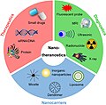 Nano Theranostics.jpg 600 × 589; 99 KByte
-
 Nichtinvasive kardiale Bildgebung.jpg 833 × 1 175; 179 KByte
-
 Nl petct.jpg 995 × 984; 129 KByte
-
 Nuclear Medicine Ir1.jpg 232 × 134; 12 KByte
-
 Nuclear Medicine Ir10.jpg 304 × 296; 19 KByte
-
 Nuclear Medicine Ir11.jpg 547 × 296; 28 KByte
-
 Nuclear Medicine Ir3.jpg 309 × 300; 19 KByte
-
 Nuclear Medicine Ir5.jpg 491 × 300; 28 KByte
-
 Nuclear Medicine Ir6.jpg 201 × 300; 14 KByte
-
 Nuclear Medicine Ir7.jpg 201 × 300; 13 KByte
-
 Nuclear Medicine Ir8.jpg 458 × 300; 22 KByte
-
 Nuclear Medicine Ir9.jpg 304 × 300; 19 KByte
-
 Nuclear medicine worker.svg 545 × 637; 49 KByte
-
 Nuclear Medicine.png 377 × 377; 107 KByte
-
-
 Nuclear Nursing.gif 600 × 472; 88 KByte
-
 Nuclide Generator Schematic-de.svg 750 × 1 375; 22 KByte
-
 Nukleamedizinische Onkologie.jpg 844 × 1 181; 173 KByte
-
 Open Catenary Two Compartment Model.svg 1 375 × 563; 9 KByte
-
 Open Mamillary Two Compartment Model.svg 1 125 × 938; 11 KByte
-
 Parathyroid subtraction.jpg 1 024 × 976; 104 KByte
-
 Paratiroide negativa.jpg 801 × 743; 51 KByte
-
 PET-MR fusion 2D.jpg 300 × 662; 62 KByte
-
 PlasmaClearanceInThreeCompartmentModel-de.svg 904 × 644; 91 KByte
-
 Positron Emission Mammography.jpg 388 × 426; 19 KByte
-
 Predictions of the open mamillary two compartment model.svg 820 × 580; 408 KByte
-
 Predictions of the renal clearance model-de.svg 844 × 624; 90 KByte
-
 Radioimmunotherapy schematic-ar.png 2 974 × 1 774; 1,14 MByte
-
 Radioimmunotherapy schematic.png 2 974 × 1 774; 989 KByte
-
 Radionuklidangiografie.JPG 746 × 627; 66 KByte
-
 Radiotraceur.jpg 971 × 298; 19 KByte
-
 Radon and Cancer by Cohen.GIF 328 × 368; 4 KByte
-
 RenalCancer-700MBq-HDP-SPECT-3hpi.jpg 1 012 × 503; 57 KByte
-
 RenalClearanceModel-de.svg 1 275 × 1 329; 13 KByte
-
 RenographyThreeCompartmentModelBackgroundAndRenogram-de.svg 844 × 624; 95 KByte
-
 RenographyThreeCompartmentModelCorrectedRenogram-de.svg 844 × 624; 86 KByte
-
 RenographyThreeCompartmentModelq4q5q6-de.svg 844 × 624; 111 KByte
-
-
-
 Sample size vs expected effect.jpg 1 800 × 1 659; 208 KByte
-
 Sanguinamento anteriore1.png 612 × 507; 69 KByte
-
 Sanguinamento posteriore1.png 622 × 533; 68 KByte
-
 SAP scan response to chemotherapy.jpg 537 × 249; 15 KByte
-
 SD-Sono001-Nummern.jpg 1 180 × 847; 631 KByte
-
 Secular eq.png 615 × 605; 72 KByte
-
 Sentinel lymph node (axilla).jpg 3 008 × 2 000; 3,41 MByte
-
 Serie Radiojodtherapie Basedow.jpg 1 515 × 484; 195 KByte
-
 Single Compartment Model.svg 375 × 750; 5 KByte
-
 SPECT CT fusion.jpg 487 × 544; 17 KByte
-
 SPECT Sinogram 360.jpg 227 × 287; 10 KByte
-
 SPECT Slice of Brain using Tc-99m Ceretec.jpg 128 × 128; 2 KByte
-
 Strahlenbelastungen.png 900 × 600; 29 KByte
-
 Stylized Phantom - Computerized Anatomical Man (CAM).jpg 444 × 254; 11 KByte
-
 Stylized Phantom - MIRD Adam and Eva.jpg 183 × 301; 11 KByte
-
 Stylized Phantom - Various Ages.jpg 381 × 278; 12 KByte
-
 Syringe Shield used in injecting Radioactive Isotope for Bone Scan.jpg 5 616 × 3 744; 17,54 MByte
-
 Taipei Veterans General Hospital Heavy Ion Therapy Center 2023-05-15 03.jpg 2 800 × 1 867; 4,52 MByte
-
 Technetium-99m Generator Tc-99m-Conetend verus Time - 2.svg 600 × 480; 14 KByte
-
 The Japaneese Society of Nuclear Medicine august 1989.jpg 2 426 × 1 889; 1,19 MByte
-
 Thorium-kuh-2.PNG 705 × 585; 18 KByte
-
 Transient eq.png 621 × 603; 75 KByte
-
 Two medical technologists use a gamma counter to measure hormone concentrations in the blood.tif 2 336 × 3 472; 13,83 MByte
-
 TwoCompartmentClosedSystemTracerTimeEvolution.svg 820 × 580; 109 KByte
-
 TwoCompartmentOpenSystemTracerTimeEvolution.svg 820 × 580; 82 KByte
-
 ⁹⁰Y-DOTATOC Structure.jpg 600 × 611; 56 KByte


.png)


.png)


.jpg)


.jpg)


