Category:Peptides
Przejdź do nawigacji
Przejdź do wyszukiwania
każdy związek chemiczny powstały z cząsteczek przynajmniej dwóch aminokwasów połączonych wiązaniem peptydowym | |||||
| Prześlij plik multimedialny | |||||
| Jest to |
| ||||
|---|---|---|---|---|---|
| Podklasa dla | |||||
| Część |
| ||||
| Składa się z | |||||
| |||||
Podkategorie
Poniżej wyświetlono 43 spośród wszystkich 43 podkategorii tej kategorii.
*
A
- Alitame (4 pliki)
- Amyloid beta-peptides (14 plików)
- Atosiban (2 pliki)
B
C
- C-peptide (1 plik)
- Cell-penetrating peptides (12 plików)
- Cetrorelix (3 pliki)
D
- Desmopressin (2 pliki)
- Dolastatins (12 plików)
E
- Echinocandin B (4 pliki)
- Elamipretide (3 pliki)
F
G
- Goserelin (3 pliki)
H
- Hirudin (9 plików)
- Histidine containing peptides (1 plik)
I
L
- Leuprorelin (4 pliki)
- Linaclotide (5 plików)
N
- Nesiritide (2 pliki)
O
P
- Peptide nucleic acids (5 plików)
- Peptidomimetics (6 plików)
S
- Silaffins (5 plików)
T
- Thiostrepton (5 plików)
V
Pliki w kategorii „Peptides”
Poniżej wyświetlono 200 spośród wszystkich 314 plików w tej kategorii.
(poprzednia strona) (następna strona)-
De-Peptid.ogg 1,8 s; 18 KB
-
LL-Q150 (fra)-WikiLucas00-peptide.wav 1,1 s; 107 KB
-
Nl-peptide.ogg 1,1 s; 14 KB
-
Pl-peptyd.ogg 1,5 s; 18 KB
-
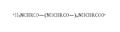
 120-154 aa that constitut Orf3b protein.png 1920 × 236; 50 KB
-
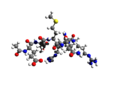
 3D view Nonatide.gif 800 × 745; 194 KB
-
 60-120 aa that constitut orf3b protein.png 1920 × 135; 32 KB
-
 7-Aminoactinomycin D.png 1282 × 731; 29 KB
-
 A polypeptide 139406160955 2.PNG 727 × 191; 5 KB
-
 A polypeptide 139406160955.PNG 683 × 214; 6 KB
-
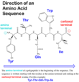 Aasequencedirection.png 511 × 527; 112 KB
-
 ABT-510.svg 1855 × 555; 78 KB
-
 Acetyl hexapeptyde-3 3D.png 800 × 600; 100 KB
-
 ACTX spider toxin 1QDP.png 2180 × 1492; 1000 KB
-
 Adegramotide.svg 1940 × 900; 192 KB
-
 Alamandine.svg 1395 × 590; 44 KB
-
 Alenix.jpg 717 × 183; 19 KB
-
 Alpha sheet bonding schematic-color.svg 2138 × 2181; 50 KB
-
 Alpha-Bungarotoxin 1IDI.png 1000 × 1133; 300 KB
-
 Alpha-Conotoxin from Conus pennaceus 1AKG.png 1000 × 1033; 180 KB
-
 Alsactide.png 2335 × 1490; 56 KB
-
-
 Analoogpeptiid.JPG 699 × 142; 34 KB
-
 Anaritide Tertiary Structure, PubChem.png 500 × 500; 12 KB
-
 Angiotensinamide.svg 512 × 516; 95 KB
-
 Antipain.svg 512 × 200; 41 KB
-
 Argireline.png 2318 × 1233; 38 KB
-
 Azagly-nafarelin.svg 3775 × 1400; 96 KB
-
 Barrel.png 1512 × 630; 67 KB
-
 Beefy meaty peptide.svg 512 × 184; 87 KB
-
 Beta sheet bonding antiparallel-color.svg 1151 × 3855; 15 KB
-
 Beta sheet bonding parallel-color.svg 1151 × 3855; 49 KB
-
 Beta-alanineVSalpha-alanine.png 530 × 335; 10 KB
-
 Beta-Chasmorphin-7 V4 transparent.svg 780 × 326; 14 KB
-
 Beta-endorphin 1-9.png 3309 × 1038; 44 KB
-
 Beta-hairpin peptide.png 1141 × 896; 79 KB
-
 Beta-peptides.png 963 × 723; 21 KB
-
 Beta-Pompilidotoxin.svg 2680 × 755; 130 KB
-
 Bioregulation.png 464 × 295; 150 KB
-
 Biphalin molecule ball.png 2123 × 1000; 443 KB
-
 Biphalin molecule spacefill.png 2000 × 1048; 510 KB
-
 Biphalin.svg 512 × 170; 81 KB
-
 Bivalirudin.png 3132 × 1211; 59 KB
-
 Bovine β-casomorphin 7.png 491 × 303; 37 KB
-
 Bulevirtide.svg 737 × 876; 177 KB
-
 Buserelin.svg 896 × 1004; 70 KB
-
 C-telopeptide.svg 1740 × 630; 46 KB
-
 C-terminal of protein.png 365 × 136; 2 KB
-
 C-terminus.svg 620 × 335; 8 KB
-
 Capreomycin IA.svg 1177 × 653; 16 KB
-
 Capreomycin.svg 2904 × 1649; 31 KB
-
 CB0184076.gif 1940 × 804; 33 KB
-
 CEND-1 aka iRGD aka LSTA1 from SMILES.tif 3230 × 3091; 226 KB
-
 CHP mouse embryo image.jpg 1779 × 1471; 608 KB
-
 CHP strand schematic.jpg 898 × 604; 126 KB
-
 Chélation.jpg 455 × 218; 19 KB
-
 Cicle 3 i 4.jpg 250 × 278; 35 KB
-
 Cilengitide.svg 1943 × 1329; 5 KB
-
 Ciprokiren skeletal.svg 374 × 196; 55 KB
-
 Cis trans isomerization kinetics X Pro peptide bonds.png 704 × 310; 7 KB
-
 Coiledcoil-wheelcartoon.png 929 × 555; 38 KB
-
 Complexe cuivre peptide.JPG 373 × 307; 9 KB
-
 Corticorelin.png 2886 × 2822; 116 KB
-
 Corticotropin sequence.svg 640 × 324; 38 KB
-
 Corticotropin-like intermediate peptide.svg 2535 × 1495; 122 KB
-
 Corticotropin.svg 5857 × 1982; 216 KB
-
 Cosyntropin.svg 1171 × 817; 158 KB
-
 Covalent linkage between CPP and nucleic aicd.jpg 960 × 720; 32 KB
-
 Cr9b00472 0009.jpg 800 × 936; 96 KB
-
 Crosslinked ultrashort peptide hydrogel.jpg 205 × 157; 33 KB
-
 D-alanine-(R)-lactate ligase.svg 571 × 207; 24 KB
-
 DDA MS Proteomics Scheme.png 907 × 491; 45 KB
-
 Decapeptide-12.svg 292 × 694; 44 KB
-
 Delphi-peptide-elpot-transparent-3D-sticks.png 1111 × 787; 124 KB
-
 Dermcidin-1L 2KSG.png 1623 × 1245; 805 KB
-
 Deslorelin.svg 2730 × 2306; 11 KB
-
 DesmopressinSek.svg 762 × 425; 109 KB
-
 Difelikefalin (2).svg 1212 × 560; 135 KB
-
 Difelikefalin.svg 2410 × 1195; 31 KB
-
 Dihexa.svg 3304 × 1278; 34 KB
-
 Direct penetrating example.png 1512 × 593; 155 KB
-
 Dirucotide.png 2365 × 902; 39 KB
-
 DL20231209-rama-final.tif 2598 × 2598; 659 KB
-
 DOTATATE.svg 2900 × 3100; 89 KB
-
 DSIP structure.png 3915 × 834; 148 KB
-
 DSIP Structure.svg 4894 × 1043; 18 KB
-
 Elobixibat skeletal.svg 437 × 222; 21 KB
-
 Emodepside.png 1024 × 1034; 23 KB
-
 Enalkiren skeletal 2.svg 324 × 256; 50 KB
-
 Enalkiren skeletal.svg 299 × 321; 49 KB
-
 Endomorphin 1.svg 512 × 275; 53 KB
-
 Endomorphin 2.svg 512 × 231; 51 KB
-
 Endomorphin-1 3D BS.png 2639 × 1518; 432 KB
-
 Enfuvirtid.svg 3816 × 362; 202 KB
-
 EnlacePeptidico-2.jpg 1500 × 500; 190 KB
-
 Enllaç peptídic.gif 960 × 576; 43 KB
-
 Esculentin-2CHa.png 419 × 452; 23 KB
-
 Estructura 2D (ɣ-MSH).png 1254 × 1438; 405 KB
-
 Estructura 2D (α-MSH).png 1426 × 1524; 501 KB
-
 Estructura 2D (β-MSH).png 1048 × 1620; 396 KB
-
 Estructura química de lantipéptido por RiPPMiner.png 558 × 406; 6 KB
-
 Etelcalcetide.svg 3901 × 1519; 60 KB
-
 Exenatide PDB=7MLL.png 1000 × 750; 284 KB
-
 Experimental hydrogel bandage.jpg 787 × 526; 241 KB
-
 Fc receptor schematic big.png 600 × 600; 10 KB
-
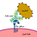 FcAr.png 600 × 600; 14 KB
-
 FE 200665.svg 512 × 273; 51 KB
-
 Feglymycin-1W7R.jpg 3858 × 1851; 834 KB
-
 Felypressin.png 1418 × 713; 27 KB
-
 Fibrinopeptide A.svg 2190 × 1485; 78 KB
-
 Fibrinopeptide B.svg 1845 × 1385; 76 KB
-
 Fibroína.png 800 × 251; 27 KB
-
 Figure 10. Copper-catalyzed cyclization of Azides.jpg 466 × 114; 11 KB
-
 Figure 11 (7834329214).png 890 × 532; 56 KB
-
-
 Figure 3 (6790806786).png 409 × 858; 385 KB
-
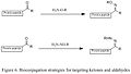
 Figure 5. Bioconjugation strategies for C-terminus.jpg 426 × 674; 25 KB
-
-
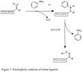 Figure 7. Nucleophilic catalysis of oxime ligation.jpg 464 × 407; 19 KB
-
 Figure 8. Staudinger Ligation with Azides.jpg 532 × 205; 14 KB
-
 Figure YYS1.svg 512 × 704; 1,97 MB
-
 Frakefamide.svg 512 × 331; 48 KB
-
 Friulimicin B.svg 2565 × 1830; 68 KB
-
 Fykoeritrin.gif 244 × 403; 3 KB
-
 GHRP-6.png 839 × 1031; 22 KB
-
 Gliadin-immuno-innate.PNG 648 × 174; 7 KB
-
 Gliadorphin-7.svg 3218 × 1215; 46 KB
-
 Glutathion oxidiert phys.svg 447 × 305; 35 KB
-
 Glyserglyalaglyala.gif 817 × 326; 4 KB
-
 Halicylindramide A,B,D,E.png 982 × 344; 15 KB
-
 Halocidin.png 378 × 398; 7 KB
-
 Hemorphin 4.svg 620 × 310; 13 KB
-
 HGH-FRAG-176-191-Chemistry.png 500 × 500; 8 KB
-
 Humangalanin.jpg 1481 × 324; 128 KB
-
 Icatibant.png 2206 × 1508; 61 KB
-
 Icatibant.svg 1890 × 830; 71 KB
-
 Ikba degron.jpg 744 × 451; 43 KB
-
 Indolicidin structure.svg 674 × 545; 35 KB
-
 Indolicidin.svg 2215 × 1175; 93 KB
-
 Integrin alphaVbeta3 and RGD Binding.png 760 × 640; 217 KB
-
 Inverted micelle1.png 1512 × 1174; 171 KB
-
 Isobaric Labeling Proteomic Workflow.png 983 × 1739; 134 KB
-
 Kallidin 3D.png 1000 × 509; 172 KB
-
 Kallidin.svg 2115 × 975; 83 KB
-
 Kassinin.png 1131 × 615; 17 KB
-
 Key steps in alternative macrocycle synthesis.png 4132 × 2568; 529 KB
-
 Key steps in bottromycin total synthesis.png 4804 × 6503; 1,37 MB
-
 L-BAPNA.svg 510 × 600; 10 KB
-
 L-Peptide-D-PeptideMirrorImages.png 2480 × 1212; 392 KB
-
 L-peptideD-peptideAnalogues.png 2055 × 2015; 875 KB
-
 Lasso-peptide-biosynthesis.png 1651 × 2229; 111 KB
-
 Lazarotide Acetato.gif 399 × 141; 2 KB
-
 LCMS Features Label Free.png 983 × 680; 110 KB
-
 Lecirelin.svg 1803 × 830; 84 KB
-
 Lepirudin sequence.svg 480 × 136; 226 KB
-
 Leu-enkaphalin.png 1742 × 550; 17 KB
-
 Leu-enkephalin molecule ball.png 2543 × 1000; 513 KB
-
 Leu-enkephalin molecule spacefill.png 2188 × 1000; 513 KB
-
 Leu-enkephalin Structure.svg 512 × 155; 126 KB
-
 Leuprorelin ball-and-stick.png 2000 × 1418; 330 KB
-
 Linusorb Fig 1.png 940 × 1362; 627 KB
-
 LY-2940094.svg 413 × 807; 6 KB
-
 MccJ25.PNG 401 × 676; 42 KB
-
 Mechanism cell uptake for CPP.jpg 1024 × 768; 172 KB
-
 Methanobactin0b3b.png 518 × 598; 123 KB
-
 Metkefamide.svg 512 × 272; 54 KB
-
 Micropeptide translation.png 2892 × 3126; 792 KB
-
 Modimelanotide.svg 7193 × 1973; 110 KB
-
 Molecular structure of peptide amphiphiles.jpg 572 × 286; 34 KB
-
 MOTS-c regulates aging metabolism and healthspan.webp 1000 × 2169; 191 KB
-
 MOTS-C.png 1544 × 398; 92 KB
-
 MRI-CPP.jpg 1019 × 533; 161 KB
-
 Muramyl dipeptide.png 919 × 474; 13 KB
-
 MVT-602 TAK-448.svg 2160 × 720; 65 KB
-
 N-terminal telopeptide.svg 1780 × 600; 46 KB
-
 N-terminus.svg 620 × 375; 8 KB
-
 Nangibotide molecular structure.png 1089 × 288; 33 KB
-
 Natriuretic Peptides and Receptors.jpg 1713 × 1133; 202 KB
-
 Neurokinin A.png 1160 × 661; 14 KB
-
 Neurokinin B.png 1141 × 555; 16 KB
-
 Neurotensin.png 3360 × 1400; 95 KB
-
 Neurotensin.svg 2495 × 820; 94 KB
-
 Nisin 2d amino structure.JPG 948 × 336; 46 KB
-
 Nitrosoglutathione.svg 512 × 221; 28 KB
-
 Nociceptin.png 1340 × 1422; 43 KB
-
 NovaPEG.jpg 1248 × 593; 39 KB
-
 Octreotide3d.png 450 × 500; 40 KB
-
 Octreotride PDB-6vc1.png 1000 × 743; 220 KB
-
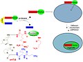
 Opening angle cpp.jpg 855 × 499; 134 KB
-
 Orf3b structure.png 1920 × 320; 67 KB
-
 Ornipressin.png 1268 × 852; 27 KB
-
 Ozarelix.svg 2004 × 804; 98 KB
-
 P53PPINetworkFromIntact.png 1280 × 1024; 1,73 MB
-
 Pal-kttks.jpg 928 × 358; 93 KB
-
 Palmitoyl pentapeptide-4.svg 3920 × 1575; 39 KB
-
 Peforelin.svg 1813 × 690; 91 KB
-
 Pegdinetanib non-peptide structure.svg 541 × 183; 39 KB
-
 Pendetide structure.svg 309 × 334; 63 KB
-
 Pentagastrin Formula V1.svg 768 × 316; 33 KB
-
 Pentagastrin.svg 1572 × 808; 40 KB


.svg)


.png)
.png)
.png)


.png)

.png)


-lactate_ligase.svg)