Category:Plant histology
Aller à la navigation
Aller à la recherche
This category include microscopic images of tissues of plants.
Sous-catégories
Cette catégorie comprend 9 sous-catégories, dont les 9 ci-dessous.
Média dans la catégorie « Plant histology »
Cette catégorie comprend 23 fichiers, dont les 23 ci-dessous.
-
 4橫切面與組織標示.png 268 × 272 ; 118 kio
-
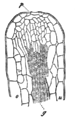 A section of hydatoda in the leaf of Primula Sinensis.png 450 × 720 ; 71 kio
-
 Bryophyta 4.png 718 × 664 ; 14 kio
-
 Cork Micrographia Hooke.png 811 × 1 170 ; 209 kio
-
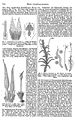
 Lacticifero de Scorzonera - enciclopedia Meyers b11 s0611.jpg 247 × 491 ; 55 kio
-
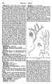
 Meyers b11 s0611.jpg 800 × 1 275 ; 428 kio
-
 Meyers b11 s0788.jpg 800 × 1 275 ; 359 kio
-
 Meyers b11 s0790.jpg 800 × 1 275 ; 347 kio
-
 Meyers b11 s0791.jpg 800 × 1 275 ; 427 kio
-
 Phloem Cells.jpg 274 × 522 ; 36 kio
-
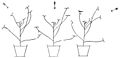
 PSM V47 D238 Downward curvature of pisum root in horizontal position.jpg 872 × 706 ; 35 kio
-
 PSM V47 D239 Separate light paths on plant leaves.jpg 1 633 × 791 ; 56 kio
-
 PSM V47 D239 Upward curvature of grass culm in horizontal position.jpg 1 352 × 1 058 ; 45 kio
-
 PSM V47 D240 Passiflora tendril after contact with a wooden rod.jpg 1 667 × 595 ; 36 kio
-
 PSM V47 D241 Longitudinal section of a tendril curvature.jpg 1 292 × 724 ; 129 kio
-
 PSM V47 D243 Curve of contraction of tendril.jpg 1 636 × 454 ; 70 kio
-
 RobertHookeMicrographia1665.jpg 1 200 × 1 600 ; 985 kio
-
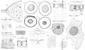 Schimper-Tafel3.jpg 2 446 × 1 440 ; 876 kio
-
 Tubos cribosos.jpg 2 824 × 2 600 ; 1,26 Mio
-
 Woody Dicot Stem- Lenticel Development in Sambucus - 36421590740 02.jpg 1 024 × 577 ; 750 kio
-
 Woody Dicot Stem- Lenticel Development in Sambucus - 36421590740 03.jpg 3 264 × 1 840 ; 4,66 Mio
-
 Woody Dicot Stem- Lenticel Development in Sambucus - 36421590740 05.jpg 3 264 × 1 840 ; 4,32 Mio
-
 Xylem Cell.jpg 231 × 467 ; 47 kio


