Category:Radiology
Naar navigatie springen
Naar zoeken springen

Français : Radiologie
Македонски: Радиологија
medisch specialisme  | |||||
| Media uploaden | |||||
| Is een | |||||
|---|---|---|---|---|---|
| Subklasse van | |||||
| Onderdeel van | |||||
| Omvat deel | |||||
| Naar verluidt hetzelfde als | Q65306418 | ||||
| |||||
Ondercategorieën
Deze categorie bevat de volgende 39 subcategorieën, van de 39 in totaal.
*
A
B
- Breast computed tomographie (3 B)
C
- Coolidge cathode tubes (11 B)
D
- Diagnostic radiology (259 B)
E
F
- Food irradiation (16 B)
G
H
I
- Images from RadsWiki (43 B)
M
N
P
R
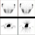
- Radionuclide imaging (11 B)
S
- SVG radiology (8 B)
T
- Tomosynthesis (2 B)
V
W
- Waste medical films (39 B)
- Whole-body irradiation (3 B)
Pagina’s in categorie "Radiology"
Deze categorie bevat de volgende 2 pagina’s, van de 2 in totaal.
Media in categorie "Radiology"
Deze categorie bevat de volgende 200 bestanden, van in totaal 274.
(vorige pagina) (volgende pagina)-
 "Health Wanted - Have Your Chest Xrayed - Find TB early" - NARA - 514453.jpg 2.141 × 3.000; 707 kB
-
 0016 ENVT Salle de Radiologie.JPG 2.950 × 1.837; 5,17 MB
-
 003 Highwood Hospital (5462834369).jpg 1.021 × 480; 359 kB
-
 003 Mansfield General (5787115720).jpg 799 × 534; 315 kB
-
 03-07-1949 05886 Röntgenapparaat (5023986238).jpg 1.024 × 811; 110 kB
-
 1911 x-ray apron.tif 1.488 × 2.845; 4,04 MB
-
 2823 Behandlingsrum för teleradium.jpg 576 × 768; 209 kB
-
 2825 Radiologi Mottagningsrum.jpg 1.024 × 751; 351 kB
-
 3847222537 378e9f1d17 bXrayBurn.jpg 2.167 × 2.831; 399 kB
-
 4408088477 e2c518947e bAnatomySchool.jpg 2.102 × 2.309; 360 kB
-
 A Coolidge X-ray tube for producing X-rays. Wellcome M0015310.jpg 4.844 × 2.233; 2,66 MB
-
 A patient being skiographed. Wellcome L0023843.jpg 1.406 × 1.434; 997 kB
-
 A poster from Liverpool's X-ray Campaign against TB Wellcome V0047918.jpg 2.362 × 3.216; 3,11 MB
-
 Accurate Reconstruction (02817002).jpg 5.568 × 3.712; 11,51 MB
-
 Acrnema1.jpg 241 × 300; 20 kB
-
 Advert for chest X-rays Wellcome L0040517.jpg 1.828 × 3.912; 1,5 MB
-
 Advert reccommending chest X-rays Wellcome L0040518.jpg 1.858 × 3.882; 1,39 MB
-
 Advertisement from the Archives of the Roentgen ray (Watson) Wellcome L0013667.jpg 1.586 × 1.190; 455 kB
-
 Advertisement from the Archives of the Roentgen ray Wellcome L0013666.jpg 1.150 × 1.629; 682 kB
-
 AGF ArctanLR.jpg 720 × 539; 145 kB
-
 American Association for Women Radiologists new logo.png 500 × 160; 18 kB
-
 Amplifier used by M. Joloiot-Curie. Wellcome M0011469.jpg 3.885 × 2.919; 1,46 MB
-

-
-
 Apparatus used by M. Joloiot-Curie. Wellcome M0011468.jpg 3.456 × 3.198; 5,62 MB
-
 Archives of physical medicine and rehabilitation (1920) (14753950081).jpg 2.020 × 2.346; 415 kB
-
 Archives of physical medicine and rehabilitation (1920) (14757172035).jpg 2.060 × 838; 195 kB
-
 Art Radiography System.jpg 5.616 × 3.744; 12,9 MB
-
 Arturo-gilardoni.png 420 × 339; 329 kB
-
 Assessing patients at the TNI field hospital (10666793275).jpg 1.440 × 956; 188 kB
-
 Aufgaben und Anforderungen an die in der Teleradiologie tätigen Personen.webp 1.654 × 1.679; 108 kB
-
 Augmentvk100 580.jpg 284 × 242; 15 kB
-
-
 Basic mechanical principles of present-day fluoroscopes.jpg 1.201 × 882; 419 kB
-
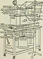
 Basic mechanical principles of radiographic apparatus.jpg 1.204 × 951; 520 kB
-
 Basic Physics of Nuclear Medicine Cassette.jpg 239 × 224; 8 kB
-
 Belize City Hospital X Ray Reading 1975.jpg 1.180 × 1.000; 961 kB
-
 Berium Bendy Straw 02.jpg 1.536 × 2.056; 1,07 MB
-
 Bone normal and degraded micro structure.jpg 735 × 739; 137 kB
-
-
 Bundesarchiv Bild 102-12853, USA, Durchleuchtung mit X-Strahlen.jpg 571 × 800; 58 kB
-
-
 Case-3-mri.png 813 × 615; 368 kB
-

-
 Childrens hospital at Annedal Gothenburg corridor.tif 2.304 × 3.307; 7,28 MB
-
 Clearly A Cyborg (5183321773).jpg 1.468 × 1.118; 145 kB
-
 Colitis-radiatio.png 440 × 435; 87 kB
-
 Collected Evidence (02817006).jpg 5.568 × 3.712; 11,34 MB
-
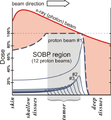 Comparison of dose profiles for proton v. x-ray radiotherapy.png 546 × 588; 54 kB
-
 Contamination Monitoring (02816998).jpg 4.629 × 3.086; 12,6 MB
-
 Control post-embolització amb coils del sagnat de l'artèria gastroduodenal.jpg 1.595 × 1.066; 210 kB
-
 Coolidge tube, United States, 1920 Wellcome L0065143.jpg 2.658 × 4.118; 604 kB
-
 Crescentsign.jpg 426 × 400; 75 kB
-
 CT Halo sign around a right lower lobe pulmonary nodule.png 512 × 204; 124 kB
-
 CT scan of a patient with Descending Necrotizing Mediastinitis.jpg 468 × 235; 37 kB
-
De-Radiologie.ogg 2,3 s; 22 kB
-

-
 Detailed Assessement (02817000).jpg 5.264 × 3.509; 10,53 MB
-
 Diagnóstico diferencial em lesões ósseas (mais de 30 anos).jpg 992 × 992; 351 kB
-
 Diagnóstico diferencial em lesões ósseas (menos de 30 anos).jpg 992 × 992; 330 kB
-

-
 DICOM InfoModel.png 601 × 571; 24 kB
-
 Dicoogle CBIR components.png 1.458 × 1.954; 574 kB
-
 Die Gartenlaube (1896) b 0276 a.jpg 1.500 × 1.018; 279 kB
-
 DirekterFlachbildDetektor.jpg 428 × 436; 19 kB
-
 Discharge tubes. Wellcome M0015829.jpg 3.500 × 3.260; 2,72 MB
-
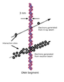 DNA-Interaction.png 816 × 1.056; 167 kB
-
 Doctor displaying x-ray.jpg 1.080 × 1.549; 244 kB
-
 Dose Calibrator Diagram.png 2.175 × 994; 129 kB
-
 DosisindikatorAnDerAnlage.jpg 4.382 × 1.958; 4,48 MB
-
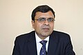 Dr. Mahaveer Prasad Goyal.jpg 3.888 × 2.592; 2,6 MB
-
 Drawing instruments (compasses, pencils) inside a case; Wellcome L0022248.jpg 1.136 × 1.704; 925 kB
-
 Dxa princip.jpg 690 × 709; 46 kB
-
 Early x-ray procedure.jpg 729 × 932; 182 kB
-
 Electrometer (quadrant type) constructed by Pierre Curie. Wellcome M0011486.jpg 1.867 × 5.896; 4,97 MB
-
 Entry to XRAY room.jpg 2.136 × 4.624; 1,33 MB
-
-
 Evidence Collection Plan (02817003).jpg 5.175 × 3.450; 9,71 MB
-
 Evidence Labels (02816995).jpg 5.362 × 3.575; 10,93 MB
-
 Evidence Recovery Team (02817004).jpg 5.568 × 3.712; 11,14 MB
-
 Evidence Recovery Team (02817005).jpg 5.316 × 3.544; 13,21 MB
-
 Experiments by Blackett in a Wilson Cloud Chamber. Wellcome M0015316.jpg 3.970 × 2.687; 4,48 MB
-
 Expositie Van Kleeftoren 2011 (6).jpg 960 × 1.280; 400 kB
-
 ExposureIndexRAD.png 754 × 675; 61 kB
-
 Falta de fusión del núcleo de la estiloides.jpg 969 × 714; 348 kB
-
 FBP Iter single.jpg 968 × 975; 43 kB
-
 First Responders (02816994).jpg 5.568 × 3.712; 10,03 MB
-
 Flowchart of the standard radiomics model.png 5.392 × 1.853; 1,37 MB
-
 Forearm internal fixation.jpg 3.456 × 4.608; 4,04 MB
-
 A Charité egyetemi kórház röntgen vizsgálója. Fortepan 74616.jpg 10.102 × 10.897; 16,11 MB
-
 Fotoleiter.jpg 285 × 288; 10 kB
-
 Fujifilm DR Calneo AQRO.jpg 3.024 × 4.032; 3,09 MB
-
 Gaiffe's Eight plate Static machine. Wellcome M0015491.jpg 3.508 × 3.025; 3,07 MB
-
 Gamma camera cross section.PNG 779 × 479; 11 kB
-
 Gedenktafel Spandauer Damm 130 (Westend) Radiologie.jpg 2.275 × 3.207; 5,83 MB
-
 Gerendertes DVT mit Nervdarstellung klein.jpg 250 × 179; 16 kB
-
 Gerendertes DVT mit Nervdarstellung.jpg 500 × 358; 19 kB
-
 Greek army medics examine X-ray, Somalia 1993.jpg 1.860 × 2.860; 3,43 MB
-
 Guantanamo captive is about to get an XRay.jpg 361 × 258; 28 kB
-
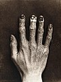
 Hand of M.H. G(lyn?); radiograph. Photograph by Sir G.P. Wellcome L0022249.jpg 1.235 × 1.642; 707 kB
-
 HCCH RCW X-ray reading.JPG 640 × 428; 52 kB
-
 Header DICOM.png 742 × 497; 19 kB
-
 Header Tag DICOM.JPG 711 × 384; 49 kB
-
 Helena Weiss, George B. Griffenhagen and Bane.jpg 630 × 508; 49 kB
-
 Histogram image against itself and spatially shifted.jpg 512 × 713; 44 kB
-
 Histograms of Bone Scan and Radiograph.jpg 660 × 201; 14 kB
-
 Histograms of pixel values in a bone scan and radiograph.gif 620 × 231; 11 kB
-
 Histograms of pixel values in a bone scan and radiograph.jpg 660 × 201; 13 kB
-
 HKMMS Bound Feet y60318.jpg 1.920 × 2.560; 1,63 MB
-
 Holder for X-ray tube in closed position. Wellcome M0015532.jpg 3.945 × 2.674; 4,53 MB
-
 Holder for X-ray tube, 1902 Wellcome M0015531.jpg 4.063 × 2.618; 3,94 MB
-
 Hopital Laquintini-4993 11.jpg 6.016 × 4.016; 7,85 MB
-
 Hopital Laquintini-4993 12.jpg 1.024 × 684; 238 kB
-
 Hopital Laquintini-4993 13.jpg 1.024 × 684; 297 kB
-
 Horizontal Impaction.jpg 3.298 × 2.638; 2,43 MB
-
 Ilisarow in Kurgan (1991).jpg 675 × 680; 221 kB
-
-
 Image Fusion Interlacing Blending.jpg 591 × 296; 23 kB
-
 IndirekterFlachbildD.jpg 932 × 582; 22 kB
-
 IndirekterFlachbildDetektor.jpg 285 × 478; 13 kB
-
 Information on X-rays impact in Bulgarian.jpg 2.240 × 4.000; 2,16 MB
-
 Internal view of amplifier used by M. Joloiot-Curie. Wellcome M0011470.jpg 3.829 × 2.924; 2,19 MB
-
 Ionisation chamber constructed and used by Pierre Curie. Wellcome M0011487.jpg 2.885 × 4.108; 1,81 MB
-
 Johannesburg Hospital, South Africa; male patient, possibly Wellcome V0029352.jpg 3.189 × 2.323; 2,71 MB
-
 Joint histograms for spatially shifted images.jpg 533 × 742; 47 kB
-
 Journal of radiology (1921) (14735299336).jpg 592 × 1.488; 112 kB
-
 Journal of radiology (1921) (14757934872).jpg 1.790 × 1.870; 265 kB
-
 Journal of radiology (1922) (14571659818).jpg 1.580 × 552; 69 kB
-
 Journal of radiology (1922) (14755091661).jpg 1.602 × 1.950; 402 kB
-
 Journal of radiology (1922) (14757911302).jpg 1.488 × 2.356; 395 kB
-
 Journal of radiology (1922) (14757948292).jpg 1.354 × 1.380; 187 kB
-
 Kerley b lines-markiert.jpg 1.891 × 1.897; 597 kB
-
 Khanh Nguyen USMC-060913-M-8770M-001.jpg 720 × 540; 62 kB
-
 X-ray treatment for a tiny sufferer. Wellcome L0015451.jpg 1.430 × 1.318; 965 kB
-
 Law Enforcement Officers (02816147).jpg 5.389 × 3.593; 10,8 MB
-
 Leone Friedman's Beauty Institute, Bucharest 1911.JPG 1.427 × 2.299; 1,02 MB
-
 Les Rayons Roentgen.jpg 603 × 898; 521 kB
-
 Limours le 25 août 2016 - 04.jpg 5.184 × 3.456; 5,35 MB
-
 Limours le 25 août 2016 - 05.jpg 5.184 × 3.456; 5,72 MB
-
 Lorgnette Humaine (2920370185).jpg 2.418 × 1.529; 748 kB
-
 Maldi ms imaging msi rat mz616.gif 432 × 159; 9 kB
-
-

-
 Marchingcubes-head.png 597 × 668; 142 kB
-
 Marta Burrel Wikimaratón de Radiología Intervencionista CIRSE 2016 - SERVEI.jpg 4.032 × 3.024; 1,85 MB
-
 Mass x-ray campaign, Liverpool 1959 (14652695395).jpg 3.072 × 2.304; 4,77 MB
-
 Mass X-ray.png 558 × 249; 152 kB
-
 Mc1.jpg 400 × 400; 46 kB
-
 Medical School-116 (6151159144).jpg 4.150 × 2.802; 6,59 MB
-
 Medizinische Panoramaaufnahme.jpg 1.574 × 902; 435 kB
-
 Military Work Dogs Require Special Care DVIDS122631.jpg 1.500 × 2.100; 1,3 MB
-
 Mineralölschutzkleidung.jpg 450 × 600; 92 kB
-
 MobiScan House.jpg 1.920 × 1.080; 945 kB
-
 Modul-electronic hg.jpg 1.131 × 781; 262 kB
-
 MS note describing the method of use of the glass tube. Wellcome M0011471.jpg 2.887 × 3.724; 1,44 MB
-
 Münster, Salzstraße 9 -- 2022 -- 4220.jpg 6.596 × 3.710; 12,92 MB
-
 Nanoprobe.jpg 550 × 397; 34 kB
-
 Nautile scanner P.JPG 1.400 × 1.240; 234 kB
-
 NM19 01.gif 386 × 202; 7 kB
-

-
 Nursers at radiology ward of Alfonso Carlos Hospital.jpg 1.602 × 2.402; 1,39 MB
-
 Only x-ray-- radium-- surgery-- ever cured cancer..JPG 566 × 768; 200 kB
-
 OrderOfAppearanceOfCarpalBones.svg 512 × 188; 73 kB
-
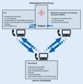
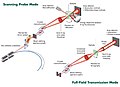
 Organigramm einer Röntgeneinrichtung.webp 2.055 × 1.842; 238 kB
-
 Original apparatus belonging to Marie Curie Wellcome M0011485.jpg 2.820 × 4.108; 1,54 MB
-
 PACS server in a radiology department.png 2.057 × 1.193; 890 kB
-
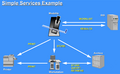 PACS-RIS Services.png 560 × 345; 66 kB
-
 Paralleltechnic-dentistry.gif 190 × 172; 4 kB
-
 Personal Protective Equipment (02816996).jpg 5.017 × 3.345; 8,76 MB
-

-
 Physikalische Grundlagen der Nuklearmedizin NM3 11.gif 489 × 402; 18 kB
-
 Pixscan II.png 553 × 338; 512 kB
-
 Plastibox , Philips rontgenapparatuur, Bestanddeelnr 903-5985.jpg 3.259 × 2.333; 1,01 MB
-
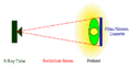
 Principe de la radiographie.png 467 × 447; 18 kB
-
 Principe radiographie faisceau divergent.png 389 × 525; 22 kB
-
 Principe radiographie.png 468 × 406; 14 kB
-
-
 Professor Gaertner experimenting with the Röntgen rays.png 2.073 × 1.773; 5,08 MB
-

-
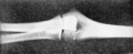 PSM V56 D0682 X ray photo of an elbow joint.png 1.626 × 666; 233 kB
-
 Rad 1300091.JPG 3.072 × 2.304; 2,89 MB
-
 Rad 1300103.JPG 2.304 × 3.072; 2,39 MB
-
 Rad 1300114.JPG 3.072 × 2.304; 2,72 MB
-
 Rad 1300120.JPG 3.072 × 2.304; 3,1 MB
-
 Rad 1300121.JPG 3.072 × 2.304; 2,34 MB
-
 Rad 1300173 cr.jpg 3.072 × 1.434; 1,55 MB
-
 Radiation Detection Equipment (02817001).jpg 5.568 × 3.712; 12,11 MB
-
 Radiation protection apron, 1920-1958 Wellcome L0065297.jpg 5.344 × 4.008; 2,78 MB
-
 Radiochromic film in hand.jpg 2.555 × 2.420; 1,49 MB
-
 Radiografia endorale digitale di una regione dentale.jpg 478 × 636; 54 kB
-
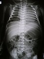
 Radiograph of the thorax. Wellcome M0013198.jpg 3.800 × 2.939; 3,04 MB
-
 Radiography and radio-therapeutics (1917) (14571426007).jpg 1.352 × 1.010; 86 kB
-
 Radiological Assessor (02816999).jpg 5.183 × 3.455; 10,45 MB
-
 Radiological Crime Scene (02816155).jpg 5.188 × 3.459; 9,58 MB
-
 Radiological exposure from medical sources.png 2.391 × 3.499; 960 kB
-
 Radiology 1904.jpg 1.707 × 1.137; 229 kB
-
 Radiology nurse 1918.png 388 × 631; 546 kB
-
 Radiology Offlice - Belize City Hospital, X-Ray Dept, 1975 - 01.jpg 5.296 × 4.140; 9,78 MB
-
 Radiology Offlice - Belize City Hospital, X-Ray Dept, 1975 - 02.jpg 5.264 × 4.166; 10,11 MB
-
 Radiology Offlice - Belize City Hospital, X-Ray Dept, 1975 - 03.jpg 3.988 × 5.335; 9,32 MB


.jpg)
.jpg)
.jpg)


.jpg)


_Wellcome_L0013667.jpg)


_(14753950081).jpg)


.jpg)


.jpg)

.jpg)


_(14777112913).jpg)
.jpg)
.jpg)
.jpg)


_b_0276_a.jpg)


_inside_a_case;_Wellcome_L0022248.jpg)


.jpg)
.jpg)
.jpg)
.jpg)

.jpg)


.jpg)


_Radiologie.jpg)


;_radiograph._Photograph_by_Sir_G.P._Wellcome_L0022249.jpg)


.jpg)
_(14741890396).jpg)


_(14757934872).jpg)
_(14755091661).jpg)
_(14757911302).jpg)
_(14757948292).jpg)


.jpg)


.jpg)


.jpg)


.jpg)


.jpg)
.jpg)


_(14758123915).jpg)


.jpg)


_(14571426007).jpg)
.jpg)
.jpg)


_(14757172035).jpg)
.png)
.jpg)
_constructed_by_Pierre_Curie._Wellcome_M0011486.jpg)
_(14735299336).jpg)
_(14571659818).jpg)
.jpg)