Category:Red blood cells
Salti al navigilo
Salti al serĉilo
most common type of blood cell  | |||||
| Alŝuti plurmedion | |||||
| Estas | |||||
|---|---|---|---|---|---|
| Subaro de | |||||
| Parto de | |||||
| Malkovrinto aŭ inventinto | |||||
| Dato de malkovro aŭ invento |
| ||||
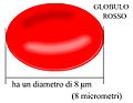
| Diametro |
| ||||
| Alia ol | |||||
| |||||
Subkategorioj
Ĉi tiu kategorio havas la 7 jenajn subkategoriojn, el 7 entute.
D
E
Dosieroj en kategorio “Red blood cells”
La jenaj 134 dosieroj estas en ĉi tiu kategorio, el 134 entute.
-
 1903 Shape of Red Blood Cells.jpg 614 × 706; 290 KB
-
 1905 Erythrocyte Life Cycle - fr.png 2 366 × 2 817; 2,02 MB
-
 1905 Erythrocyte Life Cycle - svg for text editing.svg 726 × 861; 2,6 MB
-
 1905 Erythrocyte Life Cycle.jpg 2 167 × 2 817; 1,9 MB
-
 201304 red blood cell.png 500 × 375; 31 KB
-
 A red blood cell in a capillary, pancreatic tissue - TEM.jpg 1 560 × 1 254; 579 KB
-

-
 Bacterial UTI picture of urine microscopy showing plenty of pus cells and bacteria.jpg 3 264 × 2 448; 1,07 MB
-
 Band neutrophil (16694694661).jpg 687 × 687; 491 KB
-
 Bits of Red Blood Cells under a light microscope.jpg 1 968 × 2 624; 560 KB
-
 Blausen 0761 RedBloodCells.png 1 500 × 1 500; 749 KB
-
 Blood Anemia.jpg 1 280 × 720; 187 KB
-
 Blood cell.png 1 120 × 840; 493 KB
-
 Blood cells.jpg 833 × 1 000; 74 KB
-
 Borrelia hermsii Bacteria (13758011613).jpg 3 000 × 2 250; 3,09 MB
-
 Cells in circulatory system drawing and descriptions DMHS.jpg 1 467 × 947; 152 KB
-
 Cells incirculatory system drawing and descriptions DMHSBT - May 2023.jpg 1 467 × 947; 151 KB
-
 Charged Neubauer chamber for manual blood cell counting.jpg 4 000 × 3 000; 2,34 MB
-
 Chromatin nucleofilaments.png 3 948 × 2 773; 8,24 MB
-

-
 CSIRO ScienceImage 2724 HIVpositive blood cell vs HIVnegative Blood Cell.jpg 1 772 × 1 173; 2,05 MB
-
 DHM image of human red blood cells.jpg 1 202 × 902; 60 KB
-

-

-
 Eritrocitet.jpg 1 195 × 656; 115 KB
-
 Eritrocito bio exp.jpg 319 × 360; 54 KB
-
 Eritrocito bio regulado.jpg 214 × 251; 20 KB
-
 Erythrocyte deoxy.jpg 1 280 × 800; 62 KB
-
 Erythrocyte Membrane lipids-ar.jpg 542 × 767; 88 KB
-
 Erythrocyte oxy.jpg 1 280 × 800; 58 KB
-
 Erythrocyte.png 123 × 58; 7 KB
-
 Erythrocytes (red blood cells) Rouleaux stacking.gif 440 × 440; 3,3 MB
-
 Erythrocytes in vertebrates.jpg 1 543 × 2 748; 980 KB
-
 Erythrocytes.jpg 664 × 246; 107 KB
-
 Erythropoese-ar.jpg 281 × 563; 26 KB
-
 Erythrozytopoese-1874-Koenigsberg.JPG 125 × 227; 12 KB
-
 Erytrocyte deoxy to oxy v0.6 1.gif 600 × 480; 32,15 MB
-
 Erytrocyte deoxy to oxy v0.7 AR.gif 320 × 240; 965 KB
-
 Erytrocyte deoxy to oxy v0.7.gif 320 × 240; 2,33 MB
-
 Erytrocyte deoxy to oxy v0.8 multiling labels.svg 512 × 384; 3,19 MB
-
 Erytrocyte1 951.jpg 1 280 × 1 024; 64 KB
-
 Fendo-10-00484-g003.jpg 1 084 × 437; 383 KB
-
 Fetal and Adult reticulocytes.png 921 × 346; 6 KB
-
 Globulo Rojo.png 706 × 613; 94 KB
-
 Globulorosso8micrometri.jpg 310 × 240; 31 KB
-
 GPI-SCDef.png 1 477 × 776; 530 KB
-
 Gray453-ab.png 469 × 172; 70 KB
-
 Gray453-b.png 268 × 146; 37 KB
-
 Gray453.png 469 × 305; 111 KB
-
 Größen von Partikeln in Aerosolen.png 1 748 × 1 128; 67 KB
-
 Haematopoetische farblose Blutzelle 1874.jpg 1 477 × 1 745; 517 KB
-
 Hematie.png 80 × 80; 7 KB
-
 Hematuria Microscopy.jpg 559 × 418; 36 KB
-
 Hemoglobin H disease.jpg 1 885 × 1 653; 662 KB
-
 Howell-Jolly bodies (Overview of peripheral blood stain, May-Grünwald Giemsa).jpg 2 920 × 2 940; 1,29 MB
-
 Human blood erythrocytes.jpg 1 644 × 1 040; 1,27 MB
-
 Human blood.jpg 1 600 × 1 199; 877 KB
-
 Human Cell Groups distributed by Cell Count and by Aggregate Cell Mass.jpg 3 162 × 2 096; 1,08 MB
-
 Human Cell Groups; Cell Count, Cell Mass, and Aggregate Cell Mass (Biomass).png 1 258 × 847; 149 KB
-
 Human red blood cell filmed with SFA-500 device from Biola LTD.jpg 640 × 480; 281 KB
-
 Human Red Blood Cells - Rouleau stacking.gif 440 × 440; 17,62 MB
-
 HumanRBCsPAM.png 3 500 × 2 625; 139 KB
-
 Interferogram of blood erythrocyte.jpg 709 × 678; 187 KB
-
 M. haemofelis IP2011.jpg 856 × 745; 35 KB
-
 Manual RBCs courting zone of Neubauer Chamber.jpg 4 000 × 3 000; 1,48 MB
-
 Metarubricyte.jpg 533 × 417; 143 KB
-
 Meyers b3 s0054 b1.png 327 × 363; 28 KB
-
 Micrograph of a red blood cell with basophilic stippling.jpg 319 × 306; 22 KB
-

-
 Modified sickle cell 01.jpg 378 × 291; 102 KB
-
 Morphologie érythrocytaire-1.JPG 401 × 268; 66 KB
-
 Neutrophil + monocyte (16076054013).jpg 747 × 747; 276 KB
-
 Neutrophils + monocyte (16694967012).jpg 858 × 858; 815 KB
-
 Neutrophils-hue-gray.jpg 800 × 583; 174 KB
-
 NIK 3232-Drops of blood medium.JPG 640 × 317; 15 KB
-
 Normocyte.jpg 1 141 × 1 464; 919 KB
-
 Nucleated Red Cells.jpg 1 280 × 1 024; 73 KB
-
 Observación de las células de la sangre.jpg 758 × 755; 99 KB
-
 Osmotic pressure on blood cells diagram-ar.png 1 228 × 742; 201 KB
-
 Oxygenated vs deoxygenated RBC.jpg 1 280 × 1 024; 60 KB
-
 PAM HumanRedBloodCells.png 3 500 × 2 625; 120 KB
-
 Pencil cell or Pencil shaped RBC.jpg 3 264 × 2 448; 925 KB
-
 Polychromatic erythrocyte.png 133 × 66; 9 KB
-
 Polychromophilic rbc.jpg 927 × 713; 47 KB
-
 Pus cells and RBCs in Methylene blue wet mount of pleural fluid.jpg 4 000 × 2 250; 1,68 MB
-
 Raining Red Blood Cells.jpg 4 128 × 3 096; 3,02 MB
-
 RBC membrane major proteins-ar.png 4 200 × 2 686; 3,45 MB
-
 RBC membrane major proteins.jpg 2 372 × 1 656; 733 KB
-
 RBC membrane major proteins.png 4 200 × 2 686; 3,24 MB
-
 RBC Membrane Proteins SDS-PAGE gel-ar.jpg 764 × 1 012; 134 KB
-
 RBC Membrane Proteins SDS-PAGE gel.jpg 869 × 1 013; 303 KB
-
 RBC micrograph.jpg 144 × 166; 29 KB
-
 RBC seen in neubars chamber.jpg 1 825 × 1 800; 1,5 MB
-
 Rbc's (9233940218).jpg 514 × 480; 159 KB
-
 RBC-Good.gif 170 × 240; 12 KB
-
 RBCs and Yeasts in Urine Microscopy.jpg 4 000 × 3 000; 1,16 MB
-
 RBCs, Platelets,WBC in PBS.jpg 4 000 × 2 250; 2,04 MB
-
 Red blood cell circulating in capilaries - fr.png 378 × 291; 134 KB
-
 Red blood cell diagram.PNG 512 × 384; 6 KB
-
 Red blood cell on glass.jpg 1 280 × 1 024; 57 KB
-
 Red Blood Cell.jpg 1 920 × 960; 391 KB
-
 Red blood cell.png 500 × 375; 35 KB
-
 Red blood cells (2).jpg 2 400 × 2 000; 331 KB
-
 Red blood cells (RBCs), platelets and white blood cells or leukocyte (neutrophil) on PBS.jpg 4 000 × 2 250; 1,04 MB
-
 Red blood cells by darkfield microscopy.jpg 6 000 × 4 000; 18,07 MB
-
 Red Blood Cells In Clot.png 1 023 × 696; 926 KB
-
 Red blood cells infected with malaria.jpg 1 551 × 1 229; 1,45 MB
-
 Red blood cells.jpg 1 632 × 1 224; 151 KB
-
 Red Blood Cells.jpg 2 448 × 3 264; 924 KB
-
 Red Blood Cells.png 350 × 248; 83 KB
-
 Red2.jpg 184 × 166; 17 KB
-
 Redbloodcells.jpg 144 × 166; 9 KB
-
 Rhjdm1.PNG 2 048 × 1 536; 8 MB
-
 Rhjdm2.PNG 2 048 × 1 536; 7,36 MB
-
 Rhjdm3.PNG 2 048 × 1 536; 7,56 MB
-
 Rhjdm4.PNG 2 048 × 1 536; 7,39 MB
-
 Rhjdm5.PNG 2 048 × 1 536; 7,57 MB
-
 Rouleaux formation side view.jpg 3 060 × 3 060; 1,42 MB
-
 Rubricyte.jpg 960 × 756; 387 KB
-
 Sedimented red blood cells.jpg 386 × 404; 17 KB
-
 Squeezed erythrocytes.jpg 618 × 551; 80 KB
-
 Tear Drop Cells in PBS Microscopy.jpg 3 264 × 2 448; 779 KB
-
 Thrombocytosis.jpg 2 560 × 2 048; 232 KB
-
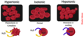 Tonicity.png 1 500 × 675; 312 KB
-

-
 Кров людини.jpg 3 000 × 2 248; 9,96 MB
-
 Невероятные эритроциты (Incredible red blood cells).jpg 3 420 × 2 243; 1,41 MB
-
 Фото № 5.jpg 1 290 × 697; 56 KB
-
 Фото № 6.jpg 720 × 887; 57 KB
-
 Фото № 7.jpg 1 432 × 802; 145 KB
-
 Эритроцит ПНГ.png 1 415 × 369; 278 KB
-
 دەڵێنە پەستان لە ناو خانەكانی خوێن بە هێڵكاری.jpg 552 × 494; 94 KB
-
 赤血球.gif 40 × 40; 2 KB
-
 赤血球浸透圧.png 554 × 288; 54 KB


.jpg)


.jpg)


_(20670817635)-ar.jpg)


_-_DPLA_-_d106dcb1bfd486d353321e358f67db49.jpg)


_Rouleaux_stacking.gif)


.jpg)


.png)


.jpg)
.jpg)


.jpg)


.jpg)
,_platelets_and_white_blood_cells_or_leukocyte_(neutrophil)_on_PBS.jpg)


_at_centre,_numerous_erythrocytes_and_platelets_(dot_like_bodies)_in_Wright%27s_stained_peripheral_blood_smear_(PBS)_microscopy.jpg)

.jpg)


