Category:Retinal ganglion cells
Vai alla navigazione
Vai alla ricerca
type of neuron located near the inner surface (ganglion cell layer) of the retina of the eye  | |||||
| Carica un file multimediale | |||||
| Istanza di | |||||
|---|---|---|---|---|---|
| Sottoclasse di |
| ||||
| |||||
File nella categoria "Retinal ganglion cells"
Questa categoria contiene 71 file, indicati di seguito, su un totale di 71.
-
 50x RGC axotomy 1 day.png 1 004 × 1 002; 680 KB
-
-
-
-
-
 Cel Ganglionar Retina Fotosens.png 964 × 303; 382 KB
-
 Cel Ganglionar Retina Fotosensible M.png 2 021 × 561; 1,01 MB
-
Coding-“What”-and-“When”-in-the-Archer-Fish-Retina-pcbi.1000977.s001.ogv 20 s, 405 × 661; 1,44 MB
-
 Color2.gif 200 × 200; 2 KB
-
Dendritic-Spikes-Amplify-the-Synaptic-Signal-to-Enhance-Detection-of-Motion-in-a-Simulation-of-the-pcbi.1000899.s001.ogv 1 min 54 s, 640 × 400; 4,72 MB
-
Developmental-patterning-of-glutamatergic-synapses-onto-retinal-ganglion-cells-1749-8104-3-8-S2.ogv 3,2 s, 1 312 × 880; 1,33 MB
-
Developmental-patterning-of-glutamatergic-synapses-onto-retinal-ganglion-cells-1749-8104-3-8-S4.ogv 19 s, 640 × 640; 1,13 MB
-
-
-
-
-
-
-
-
-
-
-
-
-
-
-
-
-
-
-
 LGN connections.gif 700 × 300; 7 KB
-
 MAX 160131Vsx1-Ath5-Draq5-ctrl3forpublic.tif (RGB)-bar.tif 2 048 × 2 048; 12 MB
-
-
-
-
-
Natural scenes through the koniocellular "Blue-on" retinal pathway.ogg 1 min 5 s, 504 × 288; 13,94 MB
-
Natural scenes through the magnocellular "OFF" retinal pathway.ogg 1 min 5 s, 504 × 288; 19,54 MB
-
Natural scenes through the magnocellular "ON" retinal pathway.ogg 1 min 5 s, 504 × 288; 18,53 MB
-
Natural scenes through the parvocellular "Green-on" retinal pathway.ogg 1 min 5 s, 504 × 288; 16,27 MB
-
Natural scenes through the parvocellular "Red-on" retinal pathway.ogg 1 min 5 s, 504 × 288; 17,59 MB
-
-
-
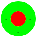
 On center off center.gif 400 × 220; 5 KB
-
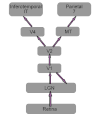 Pathway1.gif 700 × 800; 15 KB
-
Polarization-and-orientation-of-retinal-ganglion-cells-in-vivo-1749-8104-1-2-S1.ogv 15 s, 273 × 263; 1,09 MB
-
Polarization-and-orientation-of-retinal-ganglion-cells-in-vivo-1749-8104-1-2-S10.ogv 16 s, 258 × 261; 912 KB
-
Polarization-and-orientation-of-retinal-ganglion-cells-in-vivo-1749-8104-1-2-S11.ogv 10 s, 256 × 256; 804 KB
-
Polarization-and-orientation-of-retinal-ganglion-cells-in-vivo-1749-8104-1-2-S2.ogv 23 s, 124 × 283; 1,69 MB
-
Polarization-and-orientation-of-retinal-ganglion-cells-in-vivo-1749-8104-1-2-S3.ogv 7,9 s, 270 × 226; 1,24 MB
-
Polarization-and-orientation-of-retinal-ganglion-cells-in-vivo-1749-8104-1-2-S4.ogv 8,4 s, 291 × 304; 1,16 MB
-
Polarization-and-orientation-of-retinal-ganglion-cells-in-vivo-1749-8104-1-2-S5.ogv 8,9 s, 233 × 280; 384 KB
-
Polarization-and-orientation-of-retinal-ganglion-cells-in-vivo-1749-8104-1-2-S6.ogv 5,2 s, 346 × 384; 1,13 MB
-
Polarization-and-orientation-of-retinal-ganglion-cells-in-vivo-1749-8104-1-2-S7.ogv 12 s, 325 × 268; 1,18 MB
-
Polarization-and-orientation-of-retinal-ganglion-cells-in-vivo-1749-8104-1-2-S8.ogv 3,2 s, 424 × 300; 452 KB
-
Polarization-and-orientation-of-retinal-ganglion-cells-in-vivo-1749-8104-1-2-S9.ogv 16 s, 263 × 267; 1,23 MB
-
-
-
-
-
 Retina h1.jpg 1 909 × 1 200; 934 KB
-
 Retina layers1.gif 600 × 400; 46 KB
-
Retinal-Wave-Behavior-through-Activity--Dependent-Refractory-Periods-pcbi.0030245.sv001.ogv 50 s, 328 × 332; 3,63 MB
-
 Simple cell1.gif 700 × 700; 28 KB
-
-
-
Vsx2-in-the-zebrafish-retina-restricted-lineages-through-derepression-1749-8104-4-14-S1.ogv 16 s, 511 × 512; 710 KB
-
Vsx2-in-the-zebrafish-retina-restricted-lineages-through-derepression-1749-8104-4-14-S2.ogv 27 s, 407 × 345; 309 KB
-
Vsx2-in-the-zebrafish-retina-restricted-lineages-through-derepression-1749-8104-4-14-S3.ogv 32 s, 512 × 512; 3,23 MB
-
Vsx2-in-the-zebrafish-retina-restricted-lineages-through-derepression-1749-8104-4-14-S4.ogv 1 min 31 s, 480 × 360; 2,4 MB
-
Vsx2-in-the-zebrafish-retina-restricted-lineages-through-derepression-1749-8104-4-14-S5.ogv 17 s, 379 × 622; 857 KB


