Category:Scoliosis
Przejdź do nawigacji
Przejdź do wyszukiwania
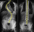
English: Scoliosis (en) is a medical condition in which a person's spine is curved from side to side, shaped like an "s", and may also be rotated.
Skrzywienie kręgosłupa  | |||||
| Prześlij plik multimedialny | |||||
| Jest to |
| ||||
|---|---|---|---|---|---|
| Podklasa dla |
| ||||
| |||||
Podkategorie
Poniżej wyświetlono 5 spośród wszystkich 5 podkategorii tej kategorii.
Pliki w kategorii „Scoliosis”
Poniżej wyświetlono 87 spośród wszystkich 87 plików w tej kategorii.
-
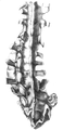
 'Part of the trunk of a crooked skeleton' Wellcome L0022317.jpg 1246 × 1722; 784 KB
-
 'Two views of the trunk of a crooked skeleton', 1733. Wellcome L0022318.jpg 1184 × 1702; 848 KB
-
 2012 01 15 0067scolio.jpg 2659 × 1337; 425 KB
-
 3D Medical Animation scoliosis Intervertibral Disc.jpg 1920 × 1080; 483 KB
-
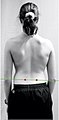
 A man suffering from Scoliosis.jpg 1267 × 3701; 577 KB
-

-
 A treatise on orthopedic surgery (1903) (14578123550).jpg 1808 × 3016; 803 KB
-
 A treatise on orthopedic surgery (1903) (14578160948).jpg 1384 × 2180; 456 KB
-
 A treatise on orthopedic surgery (1903) (14578162168).jpg 1396 × 1988; 328 KB
-
 A treatise on orthopedic surgery (1903) (14761624591).jpg 2576 × 2028; 551 KB
-
 A treatise on orthopedic surgery (1903) (14762463704).jpg 2028 × 3024; 813 KB
-
 Abnormal bone growth.jpg 353 × 509; 201 KB
-
-
 Blausen 0785 Scoliosis 01-ar.png 300 × 600; 296 KB
-
 Blausen 0785 Scoliosis 01.png 750 × 1500; 3,22 MB
-
 Boy with marked lateral curvature of the spine Wellcome L0061466.jpg 4696 × 5824; 5,82 MB
-
 Braus 1921 84.png 388 × 672; 766 KB
-
 Collage of the Abbott's method results of the bloodless scoliosis treatment.jpg 2763 × 3356; 4,52 MB
-
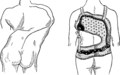
 Corset1905 210Fig186.png 1734 × 3072; 2,23 MB
-
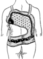
 Corset1905 211Fig187.png 3072 × 1917; 721 KB
-
 Corset1905 211Fig187a.png 1627 × 2235; 245 KB
-
 Corset1905 211Fig187b.png 1627 × 2235; 492 KB
-
 Corsetto.jpg 500 × 750; 37 KB
-
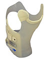 Crass Cheneau brace.jpg 513 × 681; 131 KB
-
 Different scoliosis patterns radiological and clinical highresolution.jpg 15 496 × 10 952; 11,86 MB
-
 Diseases of infancy and childhood (1914) (14772101745).jpg 2260 × 1744; 510 KB
-
 Ei 0417.jpg 530 × 450; 44 KB
-
 Esquema sección del filum terminale.png 885 × 982; 394 KB
-
 Exercises for ladies; (1836) (14584828150).jpg 3824 × 2196; 409 KB
-
 Fases en la formación de la cavidad siringomiélica.png 885 × 828; 438 KB
-
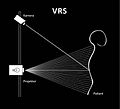 Funktionsweise VRS.jpg 454 × 413; 24 KB
-
 Gideon mantell spine1.png 473 × 872; 309 KB
-
 Gideon mantell spine2.png 422 × 873; 256 KB
-
 Gould Pyle 132.jpg 469 × 1016; 167 KB
-
 Gould Pyle 133.jpg 549 × 988; 156 KB
-
 Infantile paralysis 3.jpg 1171 × 2060; 865 KB
-
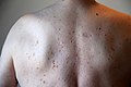 Infraspinatusatrophie.jpg 1200 × 800; 158 KB
-
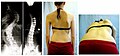
 Initial diagnosis of scoliosis with adams test and x-rays.jpg 611 × 492; 58 KB
-
 Katerina scanning new.jpg 2184 × 2076; 2 MB
-
 L. A. Sayre, Spinal disease and spinal curvature, 1877 Wellcome L0014464.jpg 1117 × 1676; 619 KB
-
 L. A. Sayre, Spinal disease and spinal curvature, 1877 Wellcome L0014466.jpg 1122 × 1707; 615 KB
-
 Lectures on orthopedic surgery (1899) (14753585886).jpg 1600 × 2460; 703 KB
-
 Leg and Arm Extensions.JPG 640 × 640; 149 KB
-
 Lewis Albert Sayre3.jpg 352 × 486; 59 KB
-
 Living anatomy and pathology; (1910) (14571495480).jpg 1752 × 2944; 511 KB
-
 Lonstein and Carlsons scoliosis progression estimation formula.svg 638 × 425; 51 KB
-
 Louis Sairy (1820 -1900) spinal deformity correction.jpg 648 × 895; 141 KB
-
 Meyers b13 s0296.jpg 800 × 1275; 312 KB
-
 Pediatrics. (1900) (14578851149).jpg 1952 × 3054; 601 KB
-
 Pediatrics. (1915) (14776406605).jpg 2976 × 1730; 443 KB
-

-
 Result after scoliosis surgery of severe scoliosis.jpg 600 × 279; 46 KB
-
 Result after scoliosis surgery.jpg 1200 × 875; 206 KB
-
 RettScoliosis.png 629 × 929; 426 KB
-
 Sayre "Spinal disease...", 1877; frontispiece Wellcome L0014467.jpg 3418 × 5199; 6,43 MB
-
 Sayre "Spinal disease...", 1877; plaster of paris bandage Wellcome L0014465.jpg 2948 × 6034; 6,53 MB
-
 Sayre "Spinal disease...", 1877; spinal deformity Wellcome L0014463.jpg 1105 × 1779; 590 KB
-
 Scoliometer, London, England, 1874-1902 Wellcome L0057973.jpg 2832 × 4256; 1,77 MB
-
 Scoliometer.jpg 420 × 200; 14 KB
-
 Scoliosi cad cam.jpg 1356 × 728; 144 KB
-
 Scoliosis brace.jpg 712 × 1445; 139 KB
-
 Scoliosis by Mishima Michiyoshi.jpg 3757 × 6997; 6,12 MB
-
 Scoliosis cobb.gif 185 × 421; 14 KB
-
 Scoliosis cobb.svg 192 × 416; 33 KB
-
 Scoliosis patient in cheneau brace correcting from 56 to 27 deg.png 1276 × 670; 1021 KB
-
 Scoliosis-mehmetnurierdem.webp 620 × 582; 41 KB
-
 Skoliometer nach Götze.jpg 2592 × 1936; 565 KB
-
 Sofia paciente.jpg 1664 × 2204; 417 KB
-
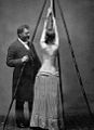
 Spinal adjustments on a scoliosis patient (1904).jpg 1896 × 1232; 416 KB
-
 Stratz Körper des Kindes 3 114.jpg 1956 × 2902; 660 KB
-
 Stratz Körper des Kindes 3 115.jpg 1956 × 2902; 787 KB
-
 Streckbett (Venel).jpg 622 × 955; 255 KB
-
 Stroop Report - Warsaw Ghetto Uprising - IPN13.jpg 1226 × 1787; 129 KB
-
 Surgical result after ventral fusion of scoliosis.jpg 1200 × 672; 132 KB
-

-
 The King In The Car Park - Page 14 - Figure 11.png 1053 × 700; 1,82 MB
-
 The King In The Car Park - Page 15 - Figure 12.png 1063 × 708; 1,88 MB
-
-
 Youth suffering from lateral spine curvature Wellcome L0062614.jpg 3653 × 5357; 2,74 MB
-

-

-
-

-
-
 Диагностическая сетка (скица).png 2400 × 6000; 267 KB
-
 Коцубан Юмейхо.jpg 900 × 1600; 129 KB
-
 Типы сколиозов по Мовшовичу.jpg 2737 × 1868; 951 KB


_(14784384802).jpg)
_(14578123550).jpg)
_(14578160948).jpg)
_(14578162168).jpg)
_(14761624591).jpg)
_(14762463704).jpg)

_(14581417359).jpg)


_(14772101745).jpg)


_(14584828150).jpg)


_(14753585886).jpg)


_(14571495480).jpg)

_spinal_deformity_correction.jpg)

_(14578851149).jpg)
_(14776406605).jpg)


.jpg)


.jpg)


_(14802013703).jpg)

_(14577245530).jpg)
_(14740917796).jpg)
_(14740934416).jpg)
.jpg)
.jpg)


_(14596927367).jpg)
.png)