Category:Single-photon emission computed tomography
Vai alla navigazione
Vai alla ricerca
English: Single-photon emission computed tomography, SPECT
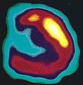
esame di medicina nucleare  Imatge SPECT de la distribució de l'examentazima de tecneci dins del cervell d'un pacient | |||||
| Carica un file multimediale | |||||
| Istanza di | |||||
|---|---|---|---|---|---|
| Sottoclasse di |
| ||||
| |||||
Sottocategorie
Questa categoria contiene le 3 sottocategorie indicate di seguito, su un totale di 3.
B
- Brain SPECT (6 F)
S
- Siemens E.CAM (1 F)
File nella categoria "Single-photon emission computed tomography"
Questa categoria contiene 56 file, indicati di seguito, su un totale di 56.
-
Activated-Platelets-in-Carotid-Artery-Thrombosis-in-Mice-Can-Be-Selectively-Targeted-with-a-pone.0018446.s001.ogv 18 s, 1 360 × 1 364; 2,79 MB
-
Activated-Platelets-in-Carotid-Artery-Thrombosis-in-Mice-Can-Be-Selectively-Targeted-with-a-pone.0018446.s002.ogv 36 s, 1 360 × 1 364; 3,04 MB
-
 Adenoviral NIS gene expression in a mouse tumour.jpg 992 × 452; 156 KB
-
Animation of a SPECT implementation using a six-axis arm robot - 2191-219X-1-32-S1.ogv 33 s, 640 × 480; 4,05 MB
-
-
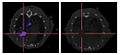
 BrainSPECT.jpg 960 × 944; 81 KB
-
 Cardiac 123I-MIBG.jpg 681 × 341; 56 KB
-
 Coded aperture mask (for gamma camera).jpg 700 × 700; 153 KB
-
-
-
 CSIRO ScienceImage 257 SPECT Camera Sinogram.jpg 2 384 × 1 528; 2,54 MB
-
 CSIRO ScienceImage 267 SPEC Diagnosis Technique.jpg 709 × 519; 299 KB
-
 Fugt-spect off peak2.jpg 9 216 × 4 450; 6,15 MB
-
 Gamma Camera SPECT Filtered Back Projection Lungs.png 236 × 256; 23 KB
-
 Gamma Camera SPECT Iterative Reconstruction Lungs.png 245 × 254; 17 KB
-
-
 Heart spect imaging.jpg 1 396 × 1 445; 779 KB
-
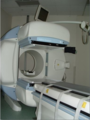 HybridSpectCTScanner.png 386 × 513; 185 KB
-
 HybridSpectCTScannerLabeled.png 385 × 512; 166 KB
-
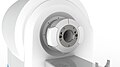 Image shows the MRI system with clip-on SPECT.JPG 2 400 × 1 350; 137 KB
-
In-Vivo-SPECT-Reporter-Gene-Imaging-of-Regulatory-T-Cells-pone.0025857.s001.ogv 9,8 s, 1 440 × 900; 2,15 MB
-
In-Vivo-SPECT-Reporter-Gene-Imaging-of-Regulatory-T-Cells-pone.0025857.s002.ogv 11 s, 1 440 × 900; 3,72 MB
-
 JASZCZAK NM phantom with flange CMH inside spect.JPG 2 448 × 3 264; 1,02 MB
-
 Low-back-pain-SPECT.jpg 472 × 332; 34 KB
-
 Mouse02-spect.gif 100 × 354; 505 KB
-
 Nuclear Medicine in Cuba (08010594) (29470523208).jpg 5 472 × 3 648; 4,03 MB
-
 Nuclear Medicine in Cuba (08010595) (28471698567).jpg 3 648 × 5 472; 2,3 MB
-
 Nuclear Medicine in Cuba (08010596) (42623325854).jpg 5 472 × 3 648; 4,95 MB
-
 Nuclear Medicine in Cuba (08010597) (29470522598).jpg 5 472 × 3 648; 4,06 MB
-
 Nuclear Medicine in Cuba (08010598) (29470521098).jpg 5 472 × 3 648; 3,95 MB
-
 Pheochromocytoma SPECT.jpg 149 × 99; 4 KB
-
Segmentation-and-Visual-Analysis-of-Whole-Body-Mouse-Skeleton-microSPECT-pone.0048976.s001.ogv 34 s, 640 × 480; 754 KB
-
Segmentation-and-Visual-Analysis-of-Whole-Body-Mouse-Skeleton-microSPECT-pone.0048976.s002.ogv 20 s, 848 × 644; 5,37 MB
-
Segmentation-and-Visual-Analysis-of-Whole-Body-Mouse-Skeleton-microSPECT-pone.0048976.s003.ogv 45 s, 1 440 × 1 080; 2,53 MB
-
Segmentation-and-Visual-Analysis-of-Whole-Body-Mouse-Skeleton-microSPECT-pone.0048976.s004.ogv 1 min 15 s, 1 440 × 1 080; 3,67 MB
-
 Small animal PET-CT-SPECT.JPG 1 110 × 849; 362 KB
-
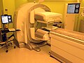
 SPECT CT OPTIMA GE 640CMH.JPG 2 448 × 3 264; 866 KB
-
 SPECT CT OPTIMA GE 641CMH.JPG 3 264 × 2 448; 932 KB
-
 SPECT CT OPTIMA GE 642CMH.JPG 3 264 × 2 448; 1,73 MB
-
 SPECT CT OPTIMA GE 643CMH.JPG 3 264 × 2 448; 1,73 MB
-
 SPECT CT OPTIMA GE 644CMH.JPG 3 264 × 2 448; 1,74 MB
-
 SPECT CT OPTIMA GE 645CMH.JPG 3 264 × 2 448; 1,75 MB
-
 SPECT CT OPTIMA GE 646CMH.JPG 3 264 × 2 448; 1,76 MB
-
 SPECT CT OPTIMA GE 647CMH.JPG 3 264 × 2 448; 1,88 MB
-
 SPECT CT OPTIMA GE 648CMH.JPG 3 264 × 2 448; 1,58 MB
-
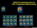 SPECT LAD.jpg 360 × 270; 68 KB
-
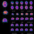
 Spect nuclear imaging slices.jpg 1 043 × 967; 1,15 MB
-
 SPECT Sinogram 360.jpg 227 × 287; 10 KB
-
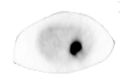
 SPECT Slice of a Liver.jpg 232 × 234; 6 KB
-
 SPECT Slice of Heart.jpg 229 × 234; 10 KB
-
 SPECT-CT of radioactive nanoparticles in mouse.png 2 059 × 2 031; 2,48 MB
-
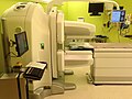
 Spectct cmh 630ge.JPG 3 264 × 2 448; 2,39 MB
-
 TORNAI-SpectrumOfMedicalImaging.jpg 720 × 504; 117 KB
-
-
 Zungengrundstruma.jpg 89 × 86; 731 byte
-
 КИД2.jpg 1 855 × 1 049; 205 KB


.jpg)


_(29470523208).jpg)
_(28471698567).jpg)
_(42623325854).jpg)
_(29470522598).jpg)
_(29470521098).jpg)