Category:Staphylococcus epidermidis
Aller à la navigation
Aller à la recherche
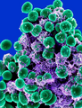
Regnum: Bacteria • Phylum: Firmicutes • Classis: Bacilli • Ordo: Bacillales • Familia: Staphylococcaceae • Genus: Staphylococcus • Species: Staphylococcus epidermidis
espèce bactérienne  Imatge d'escaneig electrònic de S. epidermidis. | |||||||||||||||||
| Téléverser des médias | |||||||||||||||||
| Nature de l’élément | |||||||||||||||||
|---|---|---|---|---|---|---|---|---|---|---|---|---|---|---|---|---|---|
| |||||||||||||||||
| |||||||||||||||||
| |||||||||||||||||
Média dans la catégorie « Staphylococcus epidermidis »
Cette catégorie comprend 43 fichiers, dont les 43 ci-dessous.
-
-
-
-
-
-
-
-
-
-
-
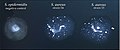
 CD8 T Cells Close to Wound After Staphylococcus Epidermidis Application.jpg 1 024 × 1 024 ; 1,11 Mio
-
 CD8 T Cells Localize Close to Wound After Staphylococcus Epidermidis Application.jpg 1 024 × 1 024 ; 930 kio
-
 Chapmanes.jpg 1 167 × 1 162 ; 396 kio
-
 Coagulase test in S. aureus and S. epidermidis.jpg 723 × 300 ; 160 kio
-
 Coagulase test.png 2 082 × 1 244 ; 2,64 Mio
-
 CoNS, S. aureus and inhibited growth of E. coli on Mannitol Salt agar (MSA).jpg 2 340 × 4 160 ; 2,32 Mio
-
 Diagnostic algorithm of possible bacterial infection.png 5 376 × 4 133 ; 3,16 Mio
-
 Gram stain of Staphylococcus epidermidis.jpg 891 × 665 ; 79 kio
-
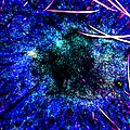 Immune Cells After Injury, Staphylococcus Epidermidis Ear Pinnae.jpg 1 000 × 1 000 ; 1,47 Mio
-
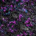 Immune Cells Surrounding Hair Follicles in Mouse Skin (7747026956).jpg 1 200 × 1 200 ; 261 kio
-
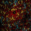 Immune Cells Surrounding Hair Follicles in Mouse Skin (7747051716).jpg 2 100 × 2 100 ; 685 kio
-
-
 Mannitol salt agar (MSA) with growth of S. aureus, CoNS and no growth of E. coli.jpg 4 000 × 2 250 ; 1,88 Mio
-
 Mannitol salt agar for Staphylococcus aureus.jpg 4 000 × 2 250 ; 2,73 Mio
-
 Mannitol Salt Agar with growth of Staphylococcus aureus and CoNS.jpg 4 000 × 2 250 ; 1,86 Mio
-
 Mannitol salt agar.jpg 2 244 × 1 798 ; 1,07 Mio
-
 MSA having growth of Staphylococcus aureus and CoNS.jpg 4 000 × 3 000 ; 5,16 Mio
-
 Rossmann-fold-1g5q.png 851 × 734 ; 233 kio
-
 S. epidermis culture.JPG 2 048 × 1 360 ; 1,34 Mio
-
 Stained Staphylococcus epidermidis.jpg 1 009 × 1 009 ; 106 kio
-
 Staphylococcus aureus (yellow colonies) and CoNS (pink colonies) on Mannitol salt agar (MSA).jpg 8 000 × 6 000 ; 9,52 Mio
-
 Staphylococcus epidermidis 01.png 700 × 412 ; 205 kio
-
 Staphylococcus epidermidis Bacteria (5613984108).jpg 492 × 640 ; 85 kio
-
 Staphylococcus epidermidis biofilm on titanium substrate.tif 2 048 × 1 536 ; 3,01 Mio
-
 Staphylococcus epidermidis colonies on Tryptic Soy Agar.jpg 1 000 × 803 ; 99 kio
-
 Staphylococcus epidermidis lores.jpg 700 × 412 ; 55 kio
-
 Staphylococcus epidermidis.jpg 1 000 × 803 ; 95 kio
-
 Staphylococcus epidermids.jpg 2 080 × 1 536 ; 2,43 Mio
-
 Staphylococcus epidermis beta-hemolytic colony on blood agar.jpg 4 000 × 3 000 ; 1,6 Mio
-
 Staphylococcus epidermis growth on blood agar.jpg 4 000 × 3 000 ; 1,37 Mio
-
 Staphylococcus epidermis.jpg 3 072 × 2 304 ; 2,37 Mio
-
 Staphylococcusepidermidis.png 492 × 640 ; 590 kio
-
 Serratia marcescens, Micrococcus luteus and Staphylococcus epidermidis.jpg 5 184 × 3 456 ; 2,87 Mio


.jpg)


.jpg)
.jpg)
_with_growth_of_S._aureus,_CoNS_and_no_growth_of_E._coli.jpg)


_and_CoNS_(pink_colonies)_on_Mannitol_salt_agar_(MSA).jpg)
.jpg)


