Category:Substantia nigra
Aller à la navigation
Aller à la recherche
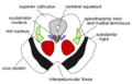
noyau du système nerveux situé au niveau du mésencéphale et du diencéphale sus-jacent, à la base des crus cerebri et ventralement par rapport au tegmentum  3D-representasjon av substantia nigra i den menneskelige hjernen. | |||||
| Téléverser des médias | |||||
| Nature de l’élément |
| ||||
|---|---|---|---|---|---|
| Sous-classe de |
| ||||
| Partie de |
| ||||
| |||||
Sous-catégories
Cette catégorie comprend seulement la sous-catégorie ci-dessous.
N
- Nigrostriatal pathway (7 F)
Média dans la catégorie « Substantia nigra »
Cette catégorie comprend 73 fichiers, dont les 73 ci-dessous.
-
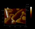
Activated-astrocytes-enhance-the-dopaminergic-differentiation-of-stem-cells-and-promote-brain-ncomms6627-s2.ogv 1 min 0 s, 1 024 × 576 ; 6,84 Mio
-
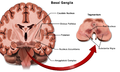
 Anatomie-Basalganglien-A.jpg 2 026 × 1 010 ; 449 kio
-
 Basal Ganglia and Related Structures-ar.svg 512 × 344 ; 48 kio
-
 Basal Ganglia and Related Structures-la.svg 523 × 352 ; 252 kio
-
 Basal Ganglia and Related Structures.svg 490 × 330 ; 39 kio
-
 Basal ganglia circuits ar.svg 1 010 × 1 453 ; 579 kio
-
 Basal ganglia circuits zh-hans.svg 1 010 × 1 453 ; 336 kio
-
 Basal ganglia circuits zh-hant.svg 1 010 × 1 453 ; 336 kio
-
 Basal ganglia circuits.svg 1 010 × 1 453 ; 341 kio
-
 Basal ganglia.svg 800 × 350 ; 76 kio
-
 Basal-ganglia-classic.png 220 × 230 ; 11 kio
-
 Basal-ganglia-coronal-sections-large.png 800 × 350 ; 122 kio
-
 Basal-ganglia-coronal-sections.png 487 × 217 ; 74 kio
-
 Blausen 0076 BasalGanglia.png 1 600 × 960 ; 1,19 Mio
-
 Blausen 0704 ParkinsonsDisease.png 2 250 × 1 350 ; 8,69 Mio
-
 Cp coronale ssthal3.jpg 589 × 534 ; 74 kio
-
 DA-loops in PD.jpg 1 050 × 834 ; 129 kio
-
 DA-loops in PD.svg 1 050 × 834 ; 84 kio
-
 Dopamine pathways -ru.svg 479 × 335 ; 27 kio
-
 Dopamine pathways ar.svg 479 × 335 ; 88 kio
-
 Dopamine Pathways vie.png 450 × 334 ; 104 kio
-
 Dopamine pathways zh-hans.svg 479 × 335 ; 27 kio
-
 Dopamine pathways zh-hant.svg 479 × 335 ; 27 kio
-
 Dopamine Pathways-es.png 500 × 371 ; 148 kio
-
 Dopamine Pathways.png 450 × 334 ; 148 kio
-
 Dopamine pathways.svg 479 × 335 ; 27 kio
-
 Dopaminergic system and reward processing.jpg 1 200 × 1 295 ; 298 kio
-
 Economo1918c.PNG 575 × 460 ; 456 kio
-
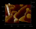 Ferritin tunneling (cropped).tif 600 × 480 ; 283 kio
-
 Ferritin tunneling.tif 704 × 576 ; 298 kio
-
 Glial cell.png 709 × 879 ; 895 kio
-
 Gliomatosis cerebri.jpg 1 200 × 1 567 ; 268 kio
-
 Gray710.png 400 × 452 ; 46 kio
-
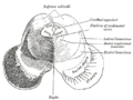 Gray711.png 500 × 373 ; 27 kio
-
 Gray712.png 500 × 321 ; 25 kio
-
 Gray717.png 500 × 568 ; 102 kio
-
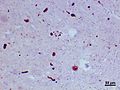
 Histological sample of Substantia nigra in Parkinson's disease.jpg 1 200 × 899 ; 263 kio
-
 Horizontal section of mibrain-superior colliculus-ja.svg 1 220 × 580 ; 146 kio
-
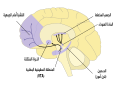
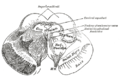
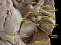
 Human brain frontal (coronal) section description 2.JPG 702 × 487 ; 43 kio
-
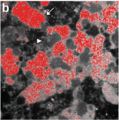 Iron outside of neuroemlanin.png 449 × 451 ; 446 kio
-
 Isodendritic core components.jpg 650 × 413 ; 214 kio
-
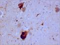
 Lewy bodies (alpha synuclein inclusions) 1.jpg 1 392 × 1 040 ; 366 kio
-
 Lewy bodies (alpha synuclein inclusions) 2.jpg 1 392 × 1 040 ; 300 kio
-
 Lewy bodies (alpha synuclein inclusions) 3.jpg 1 392 × 1 040 ; 183 kio
-
 Lewy bodies (alpha synuclein inclusions) 4.jpg 1 392 × 1 040 ; 272 kio
-
 Lewy bodies (alpha synuclein inclusions).jpg 2 800 × 2 096 ; 1,05 Mio
-
 Lewy bodies (alpha synuclein inclusions).svg 2 795 × 2 092 ; 1,48 Mio
-
 Midbraincrosssection.png 2 530 × 1 475 ; 480 kio
-
 Midbrainsuperiorcolliculus.png 357 × 229 ; 11 kio
-
 Neurogenesis and rTMS.jpg 1 200 × 551 ; 115 kio
-
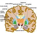
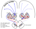
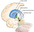 Noyau-gris-centraux3.jpg 948 × 896 ; 194 kio
-
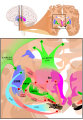
 Overview of reward structures in the human brain ar.jpg 1 200 × 1 017 ; 590 kio
-
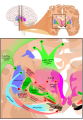
 Overview of reward structures in the human brain.jpg 1 200 × 1 017 ; 276 kio
-
-
Protection-by-the-NDI1-Gene-against-Neurodegeneration-in-a-Rotenone-Rat-Model-of-Parkinson's-Disease-pone.0001433.s004.ogv 1 min 13 s, 176 × 144 ; 394 kio
-
 Recolored Overview of reward structures in the human brain2.png 2 845 × 2 411 ; 4,9 Mio
-
 Slide10kk.JPG 960 × 720 ; 100 kio
-
 Slide2HOM.JPG 960 × 720 ; 82 kio
-
 Slide3HOM.JPG 960 × 720 ; 92 kio
-
 Substantia nigra M mulatta.tif 900 × 581 ; 1,13 Mio
-
 Substantia nigra pars compacta.jpg 451 × 377 ; 36 kio
-
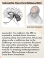 Substantia Nigra Pars Reticula Deep Brain Stimulation.png 1 728 × 2 304 ; 3 Mio
-
 Substantia nigra with Lewy body pathology.svg 2 830 × 2 117 ; 1,44 Mio
-
 Substantia nigra.gif 320 × 320 ; 608 kio
-
 Substantia Nigra.jpg 849 × 846 ; 99 kio
-
 Substantia Nigra.png 1 184 × 401 ; 352 kio
-
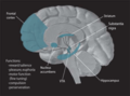
 The nuclei of the Modulatory network.png 1 267 × 1 766 ; 534 kio
-
The-temporary-and-accumulated-effects-of-transcranial-direct-current-stimulation-for-the-treatment-srep12178-s2.ogv 1 min 8 s, 320 × 240 ; 1,18 Mio
-
Ultra-High-Field-MRI-Post-Mortem-Structural-Connectivity-of-the-Human-Subthalamic-Nucleus-Video1.ogv 17 s, 686 × 602 ; 8,2 Mio
-
Vesicular-expression-and-release-of-ATP-from-dopaminergic-neurons-of-the-mouse-retina-and-midbrain-Video1.ogv 2,3 s, 512 × 512 ; 1,02 Mio
-
-
-
Vesicular-expression-and-release-of-ATP-from-dopaminergic-neurons-of-the-mouse-retina-and-midbrain-Video4.ogv 2,5 s, 512 × 512 ; 1 009 kio


_section_description_2.JPG)


_1.jpg)
_2.jpg)
_3.jpg)
_4.jpg)
.jpg)
.svg)


