Category:Temporal lobe
Vai alla navigazione
Vai alla ricerca
parte del cervello  Beyin lobları | |||||
| Carica un file multimediale | |||||
| Istanza di |
| ||||
|---|---|---|---|---|---|
| Sottoclasse di |
| ||||
| Parte di | |||||
| |||||
Sottocategorie
Questa categoria contiene le 27 sottocategorie indicate di seguito, su un totale di 27.
*
- Videos of temporal lobe (9 F)
B
- Brodmann area 20 (10 F)
- Brodmann area 21 (8 F)
- Brodmann area 22 (9 F)
- Brodmann area 34 (2 F)
- Brodmann area 36 (5 F)
- Brodmann area 37 (8 F)
- Brodmann area 38 (8 F)
- Brodmann area 41 (4 F)
- Brodmann area 42 (5 F)
- Brodmann area 52 (1 F)
C
- Collateral sulcus (11 F)
E
- Temporal lobe epilepsy (7 F)
F
H
I
- Inferior temporal gyrus (30 F)
- Inferior temporal sulcus (8 F)
M
- Middle temporal gyrus (25 F)
O
- Occipitotemporal sulcus (3 F)
P
S
- Superior temporal sulcus (24 F)
T
- Temporo-parietal junction (11 F)
W
File nella categoria "Temporal lobe"
Questa categoria contiene 37 file, indicati di seguito, su un totale di 37.
-
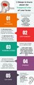 5 things to learn about the Temporal Lobe of your brain.pdf 1 250 × 3 125; 317 KB
-
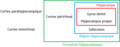 Anatomie du lobe temporal médian.png 949 × 373; 20 KB
-

-
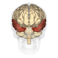
 Cerebrum - temporal lobe - anterior view.png 900 × 900; 273 KB
-
 Cerebrum - temporal lobe - inferior view.png 900 × 900; 336 KB
-
 Cerebrum - temporal lobe - lateral view.png 900 × 900; 276 KB
-
 Cerebrum - temporal lobe - posterior view.png 900 × 900; 258 KB
-

-
 Gehirn Frontalschnitt hippocampus.png 913 × 573; 263 KB
-
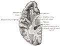
 Gray726 temporal lobe.png 992 × 573; 173 KB
-
 Gray730.png 500 × 383; 29 KB
-
 Gyri temporales transversi.png 790 × 664; 99 KB
-
 Hippolobes it.gif 400 × 261; 55 KB
-
 Hippolobes.gif 400 × 261; 56 KB
-
 Human temporal lobe areas.png 1 793 × 1 513; 1,63 MB
-
 Illu cerebrum lobes.jpg 371 × 274; 34 KB
-
 Lobus temporalis.JPG 839 × 530; 48 KB
-
 MC1 citations.pdf 1 275 × 1 650; 57 KB
-
 OccCapts.png 1 662 × 781; 847 KB
-

-
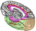
 Parahippocampal gyrus.jpg 960 × 720; 143 KB
-

-

-
 Simplified brain motor circuit.jpg 730 × 964; 55 KB
-
 Slide13ee.JPG 960 × 720; 138 KB
-
 Sobo 1909 641.png 1 027 × 730; 2,15 MB
-
 Sobo 1911 643.png 2 492 × 1 544; 11,03 MB
-
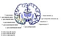
 Standard anatomical parcellation of the posterior cortical surface.png 1 810 × 1 717; 1,26 MB
-
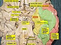
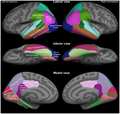
 TempCapts.png 1 899 × 856; 880 KB
-
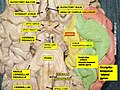
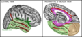
 TempCaptsLateral.png 947 × 684; 383 KB
-
 TempCaptsMedial.png 879 × 771; 425 KB
-
 Temporal lobe - anterior view.png 900 × 900; 412 KB
-
 Temporal lobe - inferior view.png 900 × 900; 449 KB
-
 Temporal lobe - inferior view2.png 900 × 900; 408 KB
-
 Temporal lobe - lateral view.png 900 × 900; 433 KB
-
 Temporal lobe - posterior view.png 900 × 900; 377 KB
-
 Tonotopic maps in human auditory cortex.jpg 1 177 × 1 280; 273 KB


