Category:Testicles
Pereiti į navigaciją
Jump to search
internal organ used in the male reproductive system  | |||||
| Įkelti mediją | |||||
| Tarimas (garso failas) | |||||
|---|---|---|---|---|---|
| Tai yra |
| ||||
| Poklasis |
| ||||
| Svarbus įvykis | |||||
| Partially coincident with |
| ||||
| |||||
English: Images and media related to testicles. See also: Category:Penis, Category:Nude men, Category:Male reproductive system
NOTE: anatomy files should be classified by species.
Subkategorijos
Rodoma 12 subkategorijų (iš viso yra 12 subkategorijų).
Daugialypės terpės rinkmenos kategorijoje „Testicles“
Rodomi 52 šios kategorijos rinkmenos (iš viso kategorijoje yra 52 rinkmenos).
-
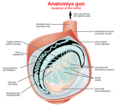 Anatomiya gun ku.png 744 × 669; 360 KiB
-
 Chickenkidneys.png 3 024 × 4 032; 15,47 MiB
-
 Chronic enlargement of the testis Wellcome L0040320.jpg 3 136 × 4 136; 2,68 MiB
-
 ControlHormFonctionAppGenMasc1.svg 1 052 × 744; 90 KiB
-
 ControlHormFonctionAppGenMasc2.svg 1 052 × 744; 128 KiB
-
-
-
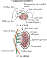 Desarrollo Testicular.png 453 × 564; 155 KiB
-
En-us-testicle.ogg 1,2 s; 15 KiB
-
 Essays and observations, physical and literary... Wellcome L0028177.jpg 1 100 × 1 678; 858 KiB
-
-
-
 Fungoid disease of the testis Wellcome L0040323.jpg 3 120 × 4 072; 2,89 MiB
-
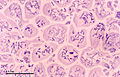
 Germinal epithelium slide.jpg 1 352 × 1 356; 175 KiB
-
 Germinal epithelium testicle.svg 410 × 360; 38 KiB
-
 GFPPupsAndTestisForWiki.jpg 1 200 × 1 704; 627 KiB
-
 Gonadenanlage1.svg 720 × 1 920; 187 KiB
-
 HHMG - linear.svg 1 060 × 1 060; 73 KiB
-
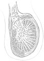
 Histology image of testis.jpg 1 800 × 4 000; 1,68 MiB
-
 Hoden-Zeichnung.jpg 700 × 963; 91 KiB
-
 Hodenschema.svg 744 × 1 052; 57 KiB
-
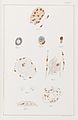 Illustration of cells infected with Leprosy bacilli Wellcome L0050078.jpg 3 879 × 5 957; 2,19 MiB
-
In-vivo-Bioimaging-as-a-Novel-Strategy-to-Detect-Doxorubicin-Induced-Damage-to-Gonadal-Blood-Vessels-pone.0023492.s003.ogv 1 min 29 s, 480 × 336; 2,73 MiB
-
In-vivo-Bioimaging-as-a-Novel-Strategy-to-Detect-Doxorubicin-Induced-Damage-to-Gonadal-Blood-Vessels-pone.0023492.s004.ogv 5 min 14 s, 480 × 336; 11,62 MiB
-
 Inflammation of the testis Wellcome L0040321.jpg 3 176 × 4 144; 2,43 MiB
-
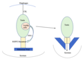 INSL3.Image.png 981 × 691; 75 KiB
-
 Internal male reproductive system in Lethocerus patruelis - ZooKeys-319-119-g001.jpeg 1 512 × 1 366; 1,9 MiB
-
Invasion-of-Wolbachia-into-Anopheles-and-Other-Insect-Germlines-in-an-Ex-vivo-Organ-Culture-System-pone.0036277.s001.ogv 3,3 s, 1 024 × 1 024; 638 KiB
-
Invasion-of-Wolbachia-into-Anopheles-and-Other-Insect-Germlines-in-an-Ex-vivo-Organ-Culture-System-pone.0036277.s002.ogv 2,1 s, 1 024 × 1 024; 219 KiB
-
-
 Meiosis (248 23).jpg 3 749 × 2 399; 2,47 MiB
-
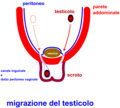 MigrazioneTesticolo.png 800 × 719; 47 KiB
-
 Mixed Germ Cell Tumor of Testis (3260625567).jpg 2 636 × 3 760; 5,17 MiB
-
 Mixed Germ Cell Tumor of Testis (w ruler) (3261449706).jpg 2 744 × 3 801; 5,63 MiB
-
Mll5-Is-Required-for-Normal-Spermatogenesis-pone.0027127.s012.ogv 33 s, 320 × 240; 1,8 MiB
-
 Normal right testis as seen on ultrasound axial view.jpg 1 024 × 768; 265 KiB
-
-
 Primary neuroendocrine tumor of testis.jpg 994 × 616; 65 KiB
-
 S16-1063 Ranochak, T MGCT- Embryonal + Seminoma + Yolk Sac.jpg 1 360 × 1 024; 329 KiB
-
 Seminiferous structure ofthe testis and epididymis Wellcome L0040319.jpg 3 150 × 4 024; 2,32 MiB
-
-
-
-
-
-
-
-
 TDS schemetic diagram.png 530 × 485; 105 KiB
-
 Testis eLife.jpg 468 × 366; 38 KiB
-
 Testis with tumours Wellcome L0040324.jpg 3 078 × 4 072; 2,49 MiB
-
 Ultrasound showing left testis calcification.jpg 1 024 × 613; 205 KiB
-
 Ultrasound showing right testis calcification.jpg 1 024 × 613; 206 KiB


.jpg)

.jpg)
_(3261449706).jpg)


