Category:Testicles
Zur Navigation springen
Zur Suche springen
männliches Geschlechtsorgan vieler Gewebetiere  | |||||
| Medium hochladen | |||||
| Utspraak as Toondatei | |||||
|---|---|---|---|---|---|
| Is en |
| ||||
| Ünnerklass vun |
| ||||
| Wichtig Begeevnis |
| ||||
| Teilweise übereinstimmend mit |
| ||||
| |||||
English: Images and media related to testicles. See also: Category:Penis, Category:Nude men, Category:Male reproductive system
NOTE: anatomy files should be classified by species.
Ünnerkategorien
De Kategorie hett disse Ünnerkategorien, vun 12 Ünnerkategorien alltohoop:
A
M
- Monorchism (0 K, 0 S, 5 D)
P
- Peritubular myoid cell (0 K, 0 S, 1 D)
R
S
T
- Testicular impact (0 K, 0 S, 19 D)
U
Mediendatein in de Kategorie „Testicles“
De Kategorie bargt disse Datein, vun 52 Datein alltohoop:
-
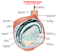 Anatomiya gun ku.png 744 × 669; 360 KB
-
 Chickenkidneys.png 3.024 × 4.032; 15,47 MB
-
 Chronic enlargement of the testis Wellcome L0040320.jpg 3.136 × 4.136; 2,68 MB
-
 ControlHormFonctionAppGenMasc1.svg 1.052 × 744; 90 KB
-
 ControlHormFonctionAppGenMasc2.svg 1.052 × 744; 128 KB
-
-
-
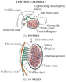 Desarrollo Testicular.png 453 × 564; 155 KB
-
En-us-testicle.ogg 1,2 s; 15 KB
-
 Essays and observations, physical and literary... Wellcome L0028177.jpg 1.100 × 1.678; 858 KB
-
-
-
 Fungoid disease of the testis Wellcome L0040323.jpg 3.120 × 4.072; 2,89 MB
-
 Germinal epithelium slide.jpg 1.352 × 1.356; 175 KB
-
 Germinal epithelium testicle.svg 410 × 360; 38 KB
-
 GFPPupsAndTestisForWiki.jpg 1.200 × 1.704; 627 KB
-
 Gonadenanlage1.svg 720 × 1.920; 187 KB
-
 HHMG - linear.svg 1.060 × 1.060; 73 KB
-
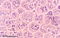
 Histology image of testis.jpg 1.800 × 4.000; 1,68 MB
-
 Hoden-Zeichnung.jpg 700 × 963; 91 KB
-
 Hodenschema.svg 744 × 1.052; 57 KB
-
 Illustration of cells infected with Leprosy bacilli Wellcome L0050078.jpg 3.879 × 5.957; 2,19 MB
-
In-vivo-Bioimaging-as-a-Novel-Strategy-to-Detect-Doxorubicin-Induced-Damage-to-Gonadal-Blood-Vessels-pone.0023492.s003.ogv 1 min 29 s, 480 × 336; 2,73 MB
-
In-vivo-Bioimaging-as-a-Novel-Strategy-to-Detect-Doxorubicin-Induced-Damage-to-Gonadal-Blood-Vessels-pone.0023492.s004.ogv 5 min 14 s, 480 × 336; 11,62 MB
-
 Inflammation of the testis Wellcome L0040321.jpg 3.176 × 4.144; 2,43 MB
-
 INSL3.Image.png 981 × 691; 75 KB
-
 Internal male reproductive system in Lethocerus patruelis - ZooKeys-319-119-g001.jpeg 1.512 × 1.366; 1,9 MB
-
-
-
-
 Meiosis (248 23).jpg 3.749 × 2.399; 2,47 MB
-
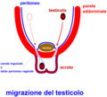 MigrazioneTesticolo.png 800 × 719; 47 KB
-
 Mixed Germ Cell Tumor of Testis (3260625567).jpg 2.636 × 3.760; 5,17 MB
-
 Mixed Germ Cell Tumor of Testis (w ruler) (3261449706).jpg 2.744 × 3.801; 5,63 MB
-
Mll5-Is-Required-for-Normal-Spermatogenesis-pone.0027127.s012.ogv 33 s, 320 × 240; 1,8 MB
-
 Normal right testis as seen on ultrasound axial view.jpg 1.024 × 768; 265 KB
-
-
 Primary neuroendocrine tumor of testis.jpg 994 × 616; 65 KB
-
 S16-1063 Ranochak, T MGCT- Embryonal + Seminoma + Yolk Sac.jpg 1.360 × 1.024; 329 KB
-
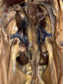
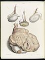
 Seminiferous structure ofthe testis and epididymis Wellcome L0040319.jpg 3.150 × 4.024; 2,32 MB
-
-
-
-
-
-
-
-
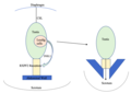
 TDS schemetic diagram.png 530 × 485; 105 KB
-
 Testis eLife.jpg 468 × 366; 38 KB
-
 Testis with tumours Wellcome L0040324.jpg 3.078 × 4.072; 2,49 MB
-
 Ultrasound showing left testis calcification.jpg 1.024 × 613; 205 KB
-
 Ultrasound showing right testis calcification.jpg 1.024 × 613; 206 KB


.jpg)

.jpg)
_(3261449706).jpg)


