Category:Virology
Przejdź do nawigacji
Przejdź do wyszukiwania
study of viruses  | |||||
| Prześlij plik multimedialny | |||||
| Jest to | |||||
|---|---|---|---|---|---|
| Podklasa dla | |||||
| |||||
This category contains media related to Virology.
Podkategorie
Poniżej wyświetlono 19 spośród wszystkich 19 podkategorii tej kategorii.
*
H
- Host antiviral responses (9 plików)
L
- Liquid nitrogen viral storage tank (4 pliki)
M
- Media from Retrovirology (27 plików)
- Media from Virology Journal (9 plików)
- Media from Viruses (12 plików)
N
P
- Plaque-Assays (9 plików)
- Polish Mykowirusy – wirusy infekujące grzyby (1 strona, 37 plików)
- Polish Wstępna charakterystyka bakteriofaga Serratia φOS10 (1 strona, 13 plików)
V
- Viral antigens (9 plików)
- Viral entry (14 plików)
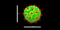
- Virion (278 plików)
- Virus-like particles (25 plików)
Pliki w kategorii „Virology”
Poniżej wyświetlono 79 spośród wszystkich 79 plików w tej kategorii.
-
De-Virologie.ogg 1,8 s; 18 KB
-
 6136 PHIL scientists PPE Ebola outbreak 1995.jpg 1024 × 667; 258 KB
-
 Adenovirus infection.jpg 1377 × 775; 137 KB
-
 AimR bound to a DNA Operator in SPbeta Phages.png 3076 × 2403; 1,13 MB
-
 Bacteriophage P22.tif 7996 × 4031; 8,04 MB
-
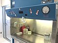 Biological Safety Cabinet (Class II, Type A2) Front view.jpg 1469 × 1102; 420 KB
-
 Biological Safety Cabinet (Class II, Type A2) Side view.jpg 1469 × 1102; 503 KB
-
 Biological Safety Cabinet (Class II, Type A2) Telstar front view.jpg 2048 × 1536; 731 KB
-
 Boonruang.jpg 400 × 500; 44 KB
-
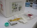
 Clinical-virus-transport-medium.jpg 3846 × 2163; 3,82 MB
-
 CP7Rgenesj.jpg 1359 × 549; 186 KB
-
 CP7Rgenesj.png 2066 × 823; 107 KB
-
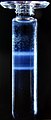 CsCl density gradient centrifugation.jpg 1217 × 2857; 2,8 MB
-
 Dengue IgM and IgG Tests-Negative.jpg 2340 × 4160; 2,43 MB
-
 Dengue RVPs.png 377 × 170; 64 KB
-
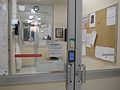 Electronic access control (BSL3 Lab).jpg 2048 × 1536; 725 KB
-
 Endogenous-Viral-Elements-in-Animal-Genomes-pgen.1001191.g004.jpg 611 × 378; 131 KB
-
-
 Graphe de la fitness d'un pathogène en fonction de la virulence.png 758 × 480; 32 KB
-
 HAV IgM and IgG Test Results.jpg 1920 × 1080; 511 KB
-
 HBsAg ELISA final reaction ready for reading.jpg 4160 × 2340; 1,03 MB
-
 HCV ELISA-Test Result.jpg 2340 × 4160; 2,45 MB
-
 HCV TRI-DOT -Positive.jpg 4160 × 2340; 2,28 MB
-
 HEV IgG Test-Positive.jpg 2340 × 4160; 2,49 MB
-
 HEV IgM and IgG Test Device.jpg 2340 × 4160; 2,38 MB
-
 History of virology.webp 5810 × 2114; 50 KB
-
 HIV ELISA final reaction in Microtiter plate wells for reading.jpg 2340 × 4160; 2,74 MB
-
 HIV Tri-Dot Test-Positive.jpg 4160 × 2340; 2,33 MB
-
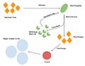
 Host virus 1.png 622 × 563; 266 KB
-
 Host virus.png 1100 × 670; 49 KB
-
 Host virus2.png 888 × 363; 91 KB
-
 Host virus3.png 506 × 426; 66 KB
-
 Host virus4.png 681 × 383; 48 KB
-
 Human virome 1.png 540 × 308; 30 KB
-
 Human virome 2 1.png 596 × 653; 299 KB
-
 Human virome.png 829 × 451; 136 KB
-
 Infektion av papillomvirus via mikroabrasioner.jpg 1012 × 514; 61 KB
-
 Livscykel papillomvirus.png 739 × 648; 105 KB
-
 Mad Scientist 4.jpg 5184 × 3456; 7,1 MB
-
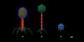
 Neutralization of SARS-CoV-2 RVPs.png 304 × 180; 40 KB
-
 Non viral vectors vs. viral vectors.png 1334 × 1056; 640 KB
-

-
 P virusollogy icon.png 500 × 450; 60 KB
-
 P4 jean mérieux.jpg 710 × 418; 87 KB
-
 PL Mykowirusy – wirusy infekujące grzyby J. Kamiński.pdf 1239 × 1754, 56 stron; 1,82 MB
-
 PL Wirusy onkolityczne. Adenowirusy w terapii przeciwnowotworowej Justyna Legierska.pdf 1239 × 1752, 65 stron; 9,6 MB
-
 PL Wstępna charakterystyka bakteriofaga Serratia φOS10 J. Kamiński.pdf 1239 × 1752, 101 stron; 2,13 MB
-
 Pre-PCR setup area.jpg 2048 × 1536; 1,58 MB
-
 Pseudotyping.jpg 720 × 960; 53 KB
-
 Receptors MV.tif 960 × 720; 132 KB
-
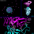 SARS-CoV-2 Infection Structural Analysis.png 7996 × 8062; 11,45 MB
-
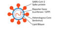 SARS-CoV-2 RVPs.png 385 × 196; 35 KB
-
 SARS-CoV-2 structural model.tiff 7996 × 4031; 92,24 MB
-
 Schéma de la coévolution.png 475 × 222; 24 KB
-
 SiRNA experiment.jpg 720 × 960; 54 KB
-
 Soluble glycoprotein production system.jpg 940 × 388; 58 KB
-
 T Bacteriophages 2024.tif 15 992 × 8063; 39,7 MB
-
 The result of the recoded VP1,2,3 of CSBV(1).png 1183 × 761; 412 KB
-
 The result of the recoded VP1,2,3 of CSBV(2).png 1175 × 771; 325 KB
-
 The result of the recoded VP1,2,3 of CSBV(3).png 1144 × 727; 164 KB
-
 The Viral Sweep.jpg 772 × 597; 39 KB
-
 ToRCH IgM Test Result.jpg 4160 × 2340; 2,25 MB
-
 Tree diagram of Pango lineages of SARS-CoV-2.svg 3469 × 3468; 1,86 MB
-
 Trinidad Regional Virus Laboratory.jpg 300 × 283; 22 KB
-
 Triple envelope packaging system for the transportation of biological samples.2.jpg 2048 × 1536; 1,56 MB
-
 Triple envelope packaging system for the transportation of biological samples.3.jpg 2048 × 1536; 1,06 MB
-
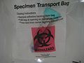 Triple envelope packaging system for the transportation of biological samples.4.jpg 2048 × 1536; 1,04 MB
-
 Triple envelope packaging system for the transportation of biological samples.jpg 2048 × 1536; 1,42 MB
-
 Typical sites of virus entry into the body in Spanish.png 607 × 600; 76 KB
-
 Typical sites of virus entry into the body.png 1500 × 1500; 346 KB
-
 Viral Phylodynamics Shankarappa.svg 988 × 720; 15 KB
-
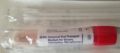 Viral transport medium.png 1154 × 510; 891 KB
-
 Virologie-Strasbourg-Microscope.jpg 2449 × 3378; 502 KB
-
 Virology 1 in 100 words - Flickr - brewbooks.jpg 1049 × 816; 114 KB
-
 VIRUS HOST.png 387 × 403; 78 KB
-
 Virus Infected Cells.jpg 1834 × 2433; 195 KB
-
 Virus Replication-zh.png 693 × 639; 152 KB
-
 Viruses-07-01902-g00S1.jpg 699 × 695; 93 KB
-
 WSV FIRST MEETING STOCKHOLM 2019.jpg 5282 × 3521; 1,95 MB


_Front_view.jpg)
_Side_view.jpg)
_Telstar_front_view.jpg)


.jpg)

.png)


.png)
.png)
.png)


