File:12985 2015 421 Fig2 HTML.webp
跳转到导航
跳转到搜索

此WEBP文件的PNG预览的大小:738 × 600像素。 其他分辨率:296 × 240像素 | 591 × 480像素 | 778 × 632像素。
原始文件 (778 × 632像素,文件大小:122 KB,MIME类型:image/webp)
说明
说明
添加一行文字以描述该文件所表现的内容
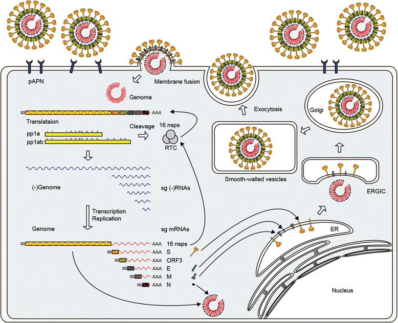
Overview of the PEDV (porcine epidemic diarrhea virus) replication cycle.
摘要
[编辑]
| 描述 |
English: Overview of the PEDV (porcine epidemic diarrhea virus) replication cycle. PEDV binds pAPN via the spike protein. Penetration and uncoating occur after the S protein-mediated fusion of the viral envelope with the plasma membrane. Following disassembly, the viral genome is released into the cytoplasm and immediately translated to yield replicases ppla and pp1ab. These polyproteins are proteolytically cleaved into 16 nsps comprising the replication and transcription complex (RTC) that first engages in the minus-strand RNA synthesis using genomic RNA. Both full- and sg-length minus strands are produced and used to synthesize full-length genomic RNA and sg mRNAs. Each sg mRNA is translated to yield only the protein encoded by the 5’-most ORF of the sg mRNA. The envelope S, E, and M proteins are inserted in the ER and anchored in the Golgi apparatus. The N protein interacts with newly synthesized genomic RNA to form helical RNP complexes. The progeny virus is assembled by budding of the preformed RNP at the ER-Golgi intermediate compartment (ERGIC) and then released by the exocytosis-like fusion of smooth-walled, virion-containing vesicles with the plasma membrane. |
| 日期 | |
| 来源 | https://virologyj.biomedcentral.com/articles/10.1186/s12985-015-0421-2 |
| 作者 | Changhee Lee |
许可协议
[编辑]
文件历史
点击某个日期/时间查看对应时刻的文件。
| 日期/时间 | 缩略图 | 大小 | 用户 | 备注 | |
|---|---|---|---|---|---|
| 当前 | 2020年7月6日 (一) 11:11 | | 778 × 632(122 KB) | Guest2625(留言 | 贡献) | Uploaded a work by Changhee Lee from https://virologyj.biomedcentral.com/articles/10.1186/s12985-015-0421-2 with UploadWizard |
您不可以覆盖此文件。
文件用途
没有页面使用本文件。
全域文件用途
以下其他wiki使用此文件:
- en.wikipedia.org上的用途
- zh.wikipedia.org上的用途