File:2011 Autophagy.tif
跳转到导航
跳转到搜索

此TIF文件的JPG预览的大小:439 × 600像素。 其他分辨率:176 × 240像素 | 351 × 480像素 | 562 × 768像素 | 749 × 1,024像素 | 1,499 × 2,048像素 | 2,609 × 3,565像素。
原始文件 (2,609 × 3,565像素,文件大小:26.65 MB,MIME类型:image/tiff)
说明
说明
添加一行文字以描述该文件所表现的内容
摘要[编辑]
| 描述 |
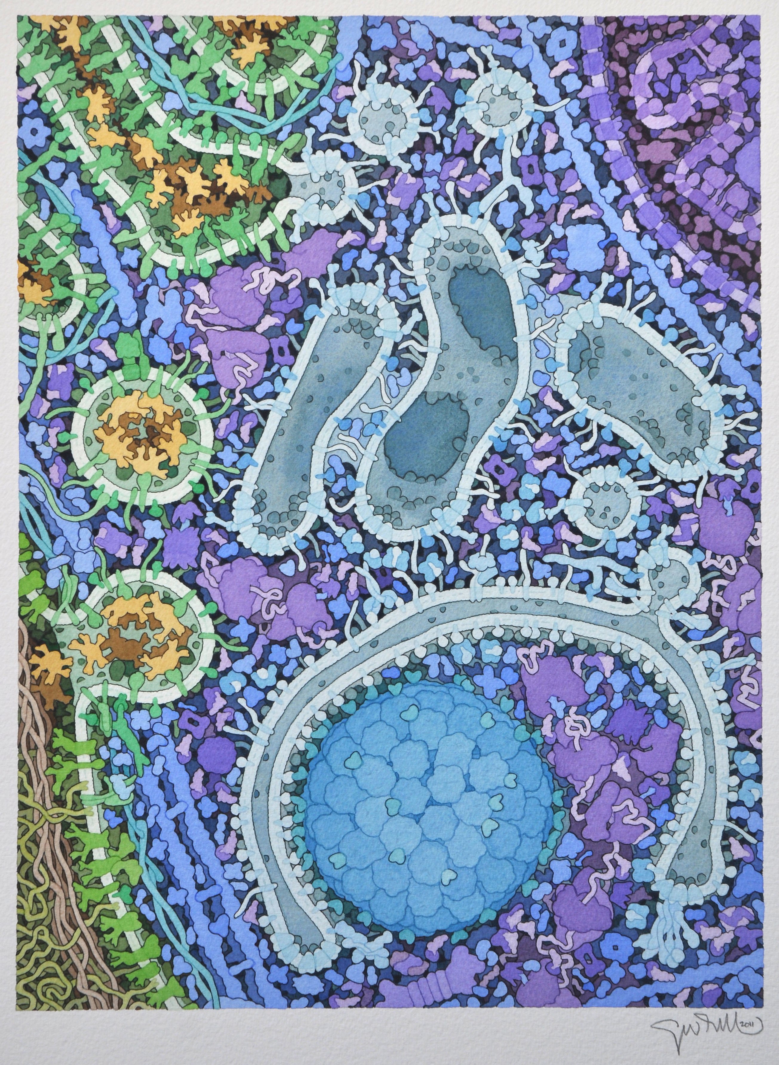
English: The formation of an autophagosome is shown, with Golgi at top left, a mitochondrion at top right, and the autophagosome at bottom center. This illustration was created in collaboration with Daniel Klionsky at the University of Michigan as a cover for the journal Cell.
For more information, see the Molecule of the Month feature on Aminopeptidase 1 and Autophagy. |
| 日期 | |
| 来源 | https://pdb101.rcsb.org/sci-art/goodsell-gallery/autophagy |
| 作者 | David Goodsell |
许可协议[编辑]
文件历史
点击某个日期/时间查看对应时刻的文件。
| 日期/时间 | 缩略图 | 大小 | 用户 | 备注 | |
|---|---|---|---|---|---|
| 当前 | 2019年7月13日 (六) 06:23 |  | 2,609 × 3,565(26.65 MB) | Evolution and evolvability(留言 | 贡献) | User created page with UploadWizard |
您不可以覆盖此文件。
文件用途
没有页面使用本文件。
全域文件用途
以下其他wiki使用此文件:
- zh.wikipedia.org上的用途