File:A computed tomography brain scan showing bilateral basal ganglia calcification.jpg
Aller à la navigation
Aller à la recherche

Taille de cet aperçu : 800 × 589 pixels. Autres résolutions : 320 × 235 pixels | 640 × 471 pixels | 1 024 × 753 pixels | 1 200 × 883 pixels.
Fichier d’origine (1 200 × 883 pixels, taille du fichier : 153 kio, type MIME : image/jpeg)
Légendes
Légendes
Ajoutez en une ligne la description de ce que représente ce fichier
| Description |
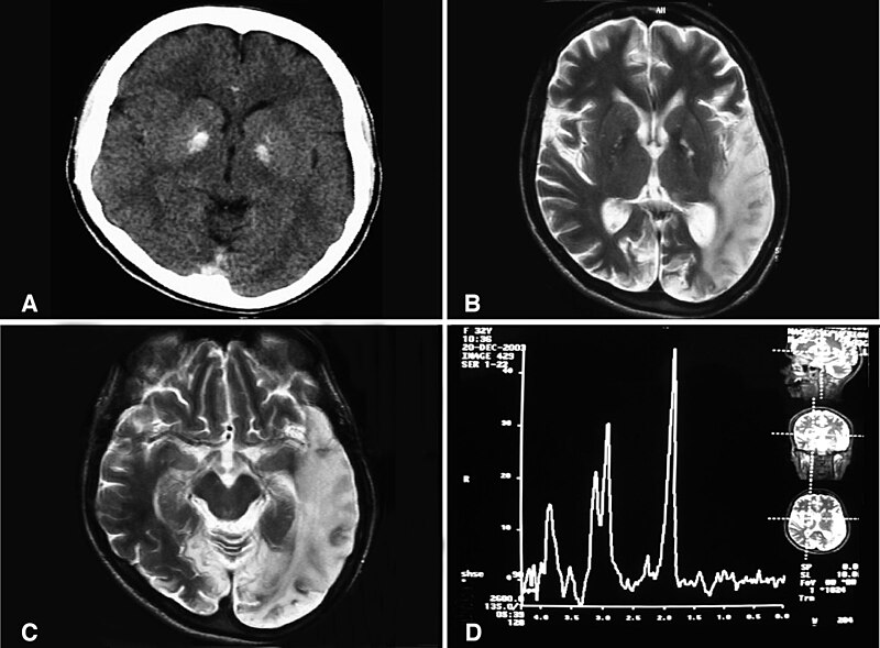
English: (a) A computed tomography brain scan showing bilateral basal ganglia calcification; the cerebellum shows prominent folia indicating mild cerebellar atrophy. (b) Axial T2 brain magnetic resonance image scan showing left temporo-parieto occipital ischemic lesion. (c) Axial T2 brain magnetic resonance image scan showing the extension of the parietal temporal region to the occipital lobe, and also showing a right occipital lesion. (d) Magnetic resonance spectroscopy showing inversion of J-coupling phenomenon at 1.3 ppm, indicating lactate peak. Abu-Amero et al. Journal of Medical Case Reports 2009 3:77 doi:10.1186/1752-1947-3-77 |
| Date | |
| Source | A patient with typical clinical features of mitochondrial encephalopathy, lactic acidosis and stroke-like episodes (MELAS) but without an obvious genetic cause: a case report |
| Auteur | Abu-Amero KK, Al-Dhalaan H, Bohlega S, Hellani A, Taylor RW. |
| Autorisation (Réutilisation de ce fichier) |
© 2009 Abu-Amero et al; licensee BioMed Central Ltd. This is an Open Access article distributed under the terms of the Creative Commons Attribution License (https://creativecommons.org/licenses/by/2.0), which permits unrestricted use, distribution, and reproduction in any medium, provided the original work is properly cited. |
Ce fichier est disponible selon les termes de la licence Creative Commons Attribution 2.0 Générique.
- Vous êtes libre :
- de partager – de copier, distribuer et transmettre cette œuvre
- d’adapter – de modifier cette œuvre
- Sous les conditions suivantes :
- paternité – Vous devez donner les informations appropriées concernant l'auteur, fournir un lien vers la licence et indiquer si des modifications ont été faites. Vous pouvez faire cela par tout moyen raisonnable, mais en aucune façon suggérant que l’auteur vous soutient ou approuve l’utilisation que vous en faites.
Historique du fichier
Cliquer sur une date et heure pour voir le fichier tel qu'il était à ce moment-là.
| Date et heure | Vignette | Dimensions | Utilisateur | Commentaire | |
|---|---|---|---|---|---|
| actuel | 28 janvier 2010 à 10:20 | | 1 200 × 883 (153 kio) | CopperKettle (d | contributions) | {{Information |Description={{en|1=(a) A computed tomography brain scan showing bilateral basal ganglia calcification; the cerebellum shows prominent folia indicating mild cerebellar atrophy. (b) Axial T2 brain magnetic resonance image scan showing left te |
Vous ne pouvez pas remplacer ce fichier.
Utilisations locales du fichier
Les 2 pages suivantes utilisent ce fichier :
Utilisations du fichier sur d’autres wikis
Les autres wikis suivants utilisent ce fichier :
- Utilisation sur ar.wikipedia.org
- Utilisation sur de.wikipedia.org
- Utilisation sur en.wikipedia.org
- Utilisation sur en.wikibooks.org
- Utilisation sur es.wikipedia.org
- Utilisation sur fa.wikipedia.org
- Utilisation sur fr.wikipedia.org
- Utilisation sur he.wikipedia.org
- Utilisation sur it.wikipedia.org
- Utilisation sur la.wikipedia.org
- Utilisation sur ru.wikipedia.org
- Utilisation sur sv.wikipedia.org
- Utilisation sur tr.wikipedia.org
- Utilisation sur www.wikidata.org
- Utilisation sur zh.wikipedia.org