File:A red blood cell in a capillary, pancreatic tissue - TEM.jpg
跳至導覽
跳至搜尋

預覽大小:746 × 600 像素。 其他解析度:299 × 240 像素 | 597 × 480 像素 | 956 × 768 像素 | 1,274 × 1,024 像素 | 1,560 × 1,254 像素。
原始檔案 (1,560 × 1,254 像素,檔案大小:579 KB,MIME 類型:image/jpeg)
說明
說明
添加單行說明來描述出檔案所代表的內容
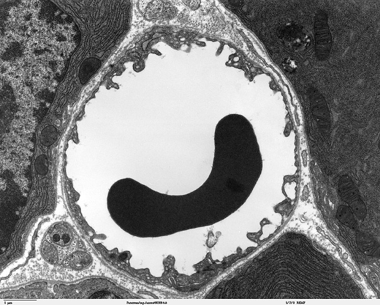
Electron microscope image of a red blood cell in a capillary in the pancreas.
摘要
[編輯]
| 描述 |
Transmission electron microscope image of a thin section cut through the pancreas(mammalian). This image shows a capillary within the pancreatic tissue(acinar cells in this image). Note the abundance of rough endoplasmic reticulum in the acinar cells. There is a red blood cell within the capillary. The capillary lining consists of long, thin endothelial cells, connected by tight junctions. The image shows fenestration of these endothelial cells. The image also shows synaptic vesicles in the neuron(nerve cell) next to the capillary. JEOL 100CX |
| 來源 | |
| 作者 | Louisa Howard |
| 授權許可 (重用此檔案) |
PD |
授權條款
[編輯]
| 此作品已由其作者,Louisa Howard,釋出至公有領域。此授權條款在全世界均適用。 這可能在某些國家不合法,如果是的話: Louisa Howard授予任何人有權利使用此作品於任何用途,除受法律約束外,不受任何限制。
|
檔案歷史
點選日期/時間以檢視該時間的檔案版本。
| 日期/時間 | 縮圖 | 尺寸 | 用戶 | 備註 | |
|---|---|---|---|---|---|
| 目前 | 2006年10月4日 (三) 23:44 | | 1,560 × 1,254(579 KB) | Patho(對話 | 貢獻) | {{Information |Description=Transmission electron microscope image of a thin section cut through the pancreas(mammalian). This image shows a capillary within the pancreatic tissue(acinar cells in this image). Note the abundance of rough endoplasmic reticul |
無法覆蓋此檔案。
檔案用途
沒有使用此檔案的頁面。
全域檔案使用狀況
以下其他 wiki 使用了這個檔案:
- bg.wikipedia.org 的使用狀況
- bn.wikipedia.org 的使用狀況
- bs.wikipedia.org 的使用狀況
- de.wikipedia.org 的使用狀況
- de.wikibooks.org 的使用狀況
- en.wikipedia.org 的使用狀況
- es.wikipedia.org 的使用狀況
- eu.wikipedia.org 的使用狀況
- fa.wikipedia.org 的使用狀況
- fr.wikipedia.org 的使用狀況
- gl.wikipedia.org 的使用狀況
- it.wikipedia.org 的使用狀況
- kk.wikipedia.org 的使用狀況
- ko.wikipedia.org 的使用狀況
- lv.wikipedia.org 的使用狀況
- mk.wikipedia.org 的使用狀況
- ml.wikipedia.org 的使用狀況
- ms.wikipedia.org 的使用狀況
- nl.wikipedia.org 的使用狀況
- nn.wikipedia.org 的使用狀況
- vi.wikipedia.org 的使用狀況
- war.wikipedia.org 的使用狀況
- www.wikidata.org 的使用狀況
- zh.wikipedia.org 的使用狀況