File:Cajal Retina.jpg
Fitxer original (500 × 745 píxels, mida del fitxer: 83 Ko, tipus MIME: image/jpeg)
Llegendes
Llegendes
Resum[modifica]
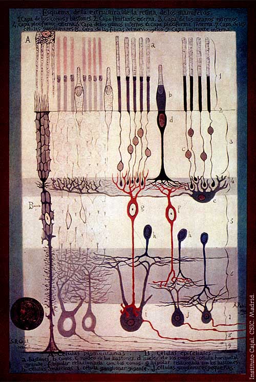
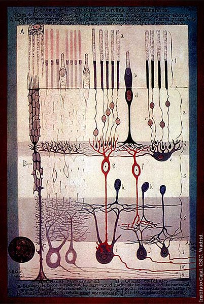
From "Structure of the Mammalian Retina" c.1900 By Santiago Ramon y Cajal.
Outline of the structure of the mammalian retina. 1. Rod and cone layer. 2. External limiting membrane. 3. Outer granular layer. 4. Outer plexiform layer. 5. Inner granular layer. 6. Inner plexiform layer. 7. Ganglion cell layer. 8. Optic nerve fibre layer. 9. Internal limiting membrane. A. Pigmented cells. B. Epithelial cells. a. Rods. b. Cones. c. Rod nucleus. d. Cone Nucleus. e. Large horizontal cell f. Cone-associated bipolar cell. g. Rod-associated bipolar cell. h. Amacrine cells. i. Giant ganglion cell. j. Small ganglion cells.
Llicència[modifica]
|
Aquest material està en domini públic als Estats Units i als altres països on el dret d'autor s'estén per 70 anys (o menys) després de la mort de l'autor.
| |
| Aquest fitxer està identificat com a lliure de restriccions conegudes sota la llei de drets d'autor, inclosos els drets veïns. | |
Historial del fitxer
Cliqueu una data/hora per veure el fitxer tal com era aleshores.
| Data/hora | Miniatura | Dimensions | Usuari/a | Comentari | |
|---|---|---|---|---|---|
| actual | 17:42, 4 març 2006 | | 500 × 745 (83 Ko) | Feezil~commonswiki (discussió | contribucions) | From "Structure of the Mammalian Retina" c.1900 By Santiago Ramon y Cajal. 1.- Rod and Cone layer 2.-Outer nuclear layer 3.- Granule layer 4.- External plexiform layer A: Pigmented cells; B: epithelial cells |
No podeu sobreescriure aquest fitxer.
Ús del fitxer
No hi ha pàgines que utilitzin aquest fitxer.
Ús global del fitxer
Utilització d'aquest fitxer en altres wikis:
- Utilització a ar.wikipedia.org
- Utilització a bn.wikipedia.org
- Utilització a ca.wikipedia.org
- Utilització a en.wikipedia.org
- Utilització a en.wikiversity.org
- Human vision and function/Part 1: How the eye works/1.3 Light stimulus and the eye
- User:Jtwsaddress42/People/Ramón y Cajal, Santiago
- User:Jtwsaddress42/People/R
- User:Jtwsaddress42/Gallery/Ramón y Cajal, Santiago
- User:Jtwsaddress42/Gallery/Ramón y Cajal, Santiago - The Visual System
- User:Jtwsaddress42/Gallery
- Utilització a es.wikipedia.org
- Utilització a et.wikipedia.org
- Utilització a ext.wikipedia.org
- Utilització a fa.wikipedia.org
- Utilització a fr.wikipedia.org
- Utilització a gl.wikipedia.org
- Utilització a he.wikipedia.org
- Utilització a hy.wikipedia.org
- Utilització a it.wikipedia.org
- Utilització a ja.wikipedia.org
- Utilització a ko.wikipedia.org
- Utilització a ml.wikipedia.org
- Utilització a pt.wikipedia.org
- Utilització a ru.wikipedia.org
- Utilització a simple.wikipedia.org
- Utilització a th.wikipedia.org
- Utilització a uk.wikipedia.org
- Utilització a vi.wikipedia.org
- Utilització a zh.wikipedia.org