File:Chlamydomonas TEM 04.jpg
ナビゲーションに移動
検索に移動

このプレビューのサイズ: 751 × 600 ピクセル。 その他の解像度: 301 × 240 ピクセル | 601 × 480 ピクセル | 961 × 768 ピクセル | 1,280 × 1,023 ピクセル | 1,800 × 1,438 ピクセル。
元のファイル (1,800 × 1,438 ピクセル、ファイルサイズ: 720キロバイト、MIME タイプ: image/jpeg)
キャプション
キャプション
このファイルの内容を1行で記述してください
| 解説 |
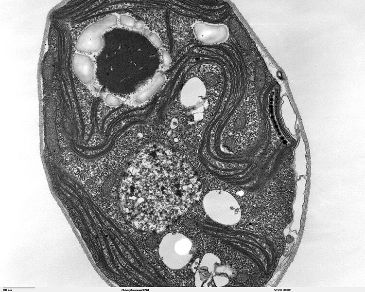
Transmission electron microscope image, showing an example of green algae (Chlorophyta). Chlamydomanas reinhardtii is a unicellular flagellate used as a model system in molecular genetics work and flagellar motility studies. This image of a thin section through a whole Chlamydomonas, shows the chloroplast, pyrenoid(surrounded by a shell of starch grains), mitochondria, nucleus, vacuoles, eye spot, and the cell wall. |
| 日付 | |
| 原典 | Source and public domain notice at http://remf.dartmouth.edu/imagesindex.html |
| 作者 | Dartmouth Electron Microscope Facility, Dartmouth College |
| 許可 (ファイルの再利用) |
Released into the public domain |
| この著作物は、著作者であるDartmouth Electron Microscope Facility, Dartmouth Collegeによって権利が放棄され、パブリックドメインとされました。これは全世界で適用されます。 一部の国では、これが法的に可能ではない場合があります。その場合は、次のように宣言します。 Dartmouth Electron Microscope Facility, Dartmouth Collegeは、あらゆる人に対して、法により必要とされている条件を除き、如何なる条件も課すことなく、あらゆる目的のためにこの著作物を使用する権利を与えます。
|
ファイルの履歴
過去の版のファイルを表示するには、その版の日時をクリックしてください。
| 日付と時刻 | サムネイル | 寸法 | 利用者 | コメント | |
|---|---|---|---|---|---|
| 現在の版 | 2007年9月21日 (金) 06:42 | | 1,800 × 1,438 (720キロバイト) | Neil916 (トーク | 投稿記録) | {{Information |Description= Transmission electron microscope image, showing an example of green algae (Chlorophyta). ''Chlamydomanas reinhardtii'' is a unicellular flagellate used as a model system in molecular genetics work and flagellar motility studie |
このファイルは上書きできません。
ファイルの使用状況
このファイルを使用しているページはありません。
グローバルなファイル使用状況
以下に挙げる他のウィキがこの画像を使っています:
- ja.wikipedia.org での使用状況