File:Chlamydomonas TEM 09.jpg
跳至導覽
跳至搜尋

預覽大小:751 × 600 像素。 其他解析度:301 × 240 像素 | 601 × 480 像素 | 961 × 768 像素 | 1,280 × 1,023 像素 | 1,800 × 1,438 像素。
原始檔案 (1,800 × 1,438 像素,檔案大小:784 KB,MIME 類型:image/jpeg)
說明
說明
添加單行說明來描述出檔案所代表的內容
| 描述 |
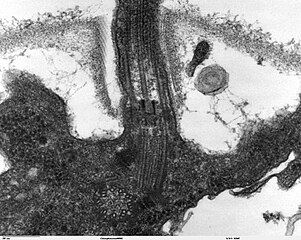
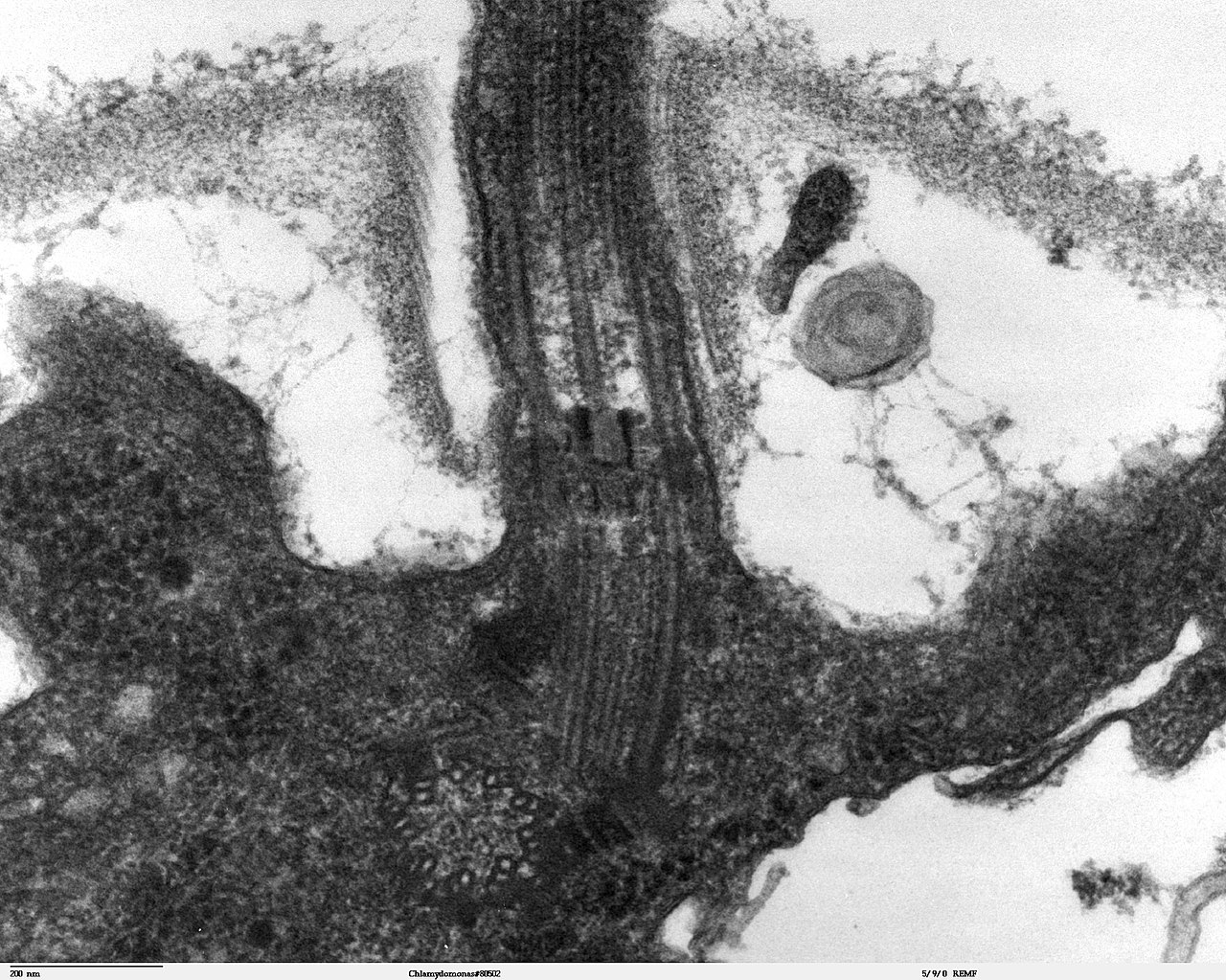
Transmission electron microscope image, showing an example of green algae (Chlorophyta). Chlamydomanas reinhardtii is a unicellular flagellate used as a model system in molecular genetics work and flagellar motility studies. This image is a longitudinal section through the flagella area. In the cell apex is the basal body that is the anchoring site for a flagella. Basal bodies originate from and have a substructure similar to that of centrioles, with nine peripheral microtubule triplets(see structure at bottom center of image). The two inner microtubules of each triplet in a basal body become the two outer doublets in the flagella. This image also shows the transition region, with its fibers of the stellate structure. The top of the image shows the flagella passing through the cell wall. |
| 日期 | |
| 來源 | Source and public domain notice at http://remf.dartmouth.edu/imagesindex.html |
| 作者 | Dartmouth Electron Microscope Facility, Dartmouth College |
| 授權許可 (重用此檔案) |
Released into the public domain |
| 此作品已由其作者,Dartmouth Electron Microscope Facility, Dartmouth College,釋出至公有領域。此授權條款在全世界均適用。 這可能在某些國家不合法,如果是的話: Dartmouth Electron Microscope Facility, Dartmouth College授予任何人有權利使用此作品於任何用途,除受法律約束外,不受任何限制。
|
檔案歷史
點選日期/時間以檢視該時間的檔案版本。
| 日期/時間 | 縮圖 | 尺寸 | 使用者 | 備註 | |
|---|---|---|---|---|---|
| 目前 | 2007年9月21日 (五) 06:47 | | 1,800 × 1,438(784 KB) | Neil916(留言 | 貢獻) | {{Information |Description= Transmission electron microscope image, showing an example of green algae (Chlorophyta). <br><br>''Chlamydomanas reinhardtii'' is a unicellular flagellate used as a model system in molecular genetics work and flagellar motilit |
無法覆蓋此檔案。
檔案用途
沒有使用此檔案的頁面。
全域檔案使用狀況
以下其他 wiki 使用了這個檔案:
- ar.wikipedia.org 的使用狀況
- bs.wikipedia.org 的使用狀況
- ca.wikipedia.org 的使用狀況
- cs.wikipedia.org 的使用狀況
- de.wikipedia.org 的使用狀況
- de.wikibooks.org 的使用狀況
- en.wikipedia.org 的使用狀況
- es.wikipedia.org 的使用狀況
- gl.wikipedia.org 的使用狀況
- id.wikipedia.org 的使用狀況
- ja.wikipedia.org 的使用狀況
- ko.wikipedia.org 的使用狀況
- pl.wikipedia.org 的使用狀況
- ru.wikipedia.org 的使用狀況
- sv.wikipedia.org 的使用狀況
- tr.wikipedia.org 的使用狀況
- uk.wikipedia.org 的使用狀況
- zh.wikipedia.org 的使用狀況