File:EB1911 Haemosporidia - life-cycle of the parasite of pernicious malaria.jpg

Faylın orijinalı (1.071 × 1.600 piksel, fayl həcmi: 500 KB, MIME növü: image/jpeg)
Captions
Captions
Xülasə
[redaktə]| İzah |
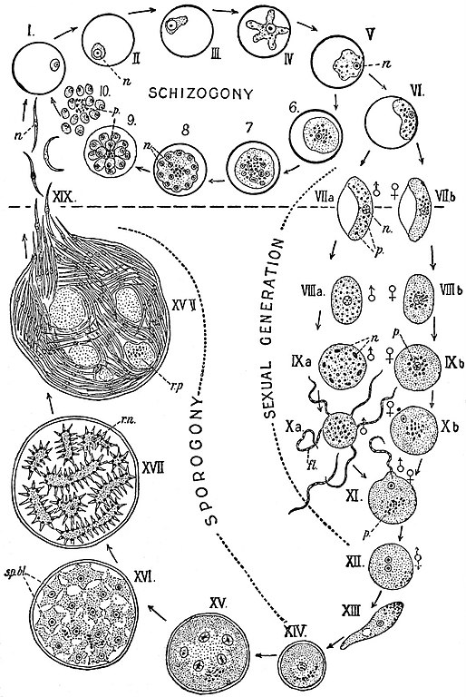
English: Diagram of the complete life-cycle of the parasite of pernicious malaria, Laverania malariae, Gr. et Fel. The stages on the upper side of the dotted line are those found in human blood; below the dotted line are seen the phases through which the parasite passes in the intermediate host, the mosquito. Plan and arrangement chiefly after Neveu-Lemaire; details of the figures founded on those of Grassi, Schaudinn (Leuckart’s Zoologische Wandtafeln), Ross and others:
I.-V. and 6-10 show the schizogony. VI.-XII., The sexual generation. XIII., The motile zygote. XIV.-XIX., Sporogony. I.-III., Young amoebulae in blood-corpuscles. IV., Older, actively amoeboid trophozoite. V., Still older, less amoeboid trophozoite. 6, Mature schizont. 7, Schizont, with nucleus dividing up. 8, Young rosette stage. 9, Fully formed rosette stage. 10, Merozoites free in the blood by breaking down of the corpuscle. VI., Young indifferent gametocyte. VII., a, Male crescent. VII., b, Female crescent. VIII., a and b, The gametocytes becoming oval. IX., a and b, Spherical gametocytes; in the male (IX. a) the nucleus has divided up. X., a and b, Formation of gametes; in the male (X. a) the so-called flagella or male gametes (fl) are thrown out, one of them is seen detached; in the female (X. b) a portion of the nucleus has been expelled. XI., A male gamete penetrating a female gamete at a cone of reception formed near the nucleus. XII., Zygote with two pronuclei in proximity. XIII., Zygote in the motile stage (vermicule or oökinete). XIV., Encysted zygote (oöcyst). XV., Commencing multiplication of the nuclei in the oöcyst. XVI., Oöcyst with numerous sporoblasts. XVII., Commencing formation of sporozoites. XVIII., Full-grown oocyst crammed with ripe sporozoites; on one side the cyst has burst and the sporozoites are escaping. XIX., Free sporozoites, showing their changes of form. n, Nucleus of the parasite. p, Melanin pigment. fl, “Flagella.” sp. bl., Sporoblasts. r. n., Residual nuclei. r. p., Residual protoplasm. |
|||
| Tarix | published 1911 | |||
| Mənbə | “Haemosporidia,” Encyclopædia Britannica (11th ed.), v. 12, 1911, p. 808, fig. 1. | |||
| Müəllif | From Lankester’s Treatise on Zoology. | |||
| İcazə (Faylın təkrar istifadəsi) |
|

Faylın tarixçəsi
Faylın əvvəlki versiyasını görmək üçün gün/tarix bölməsindəki tarixlərə klikləyin.
| Tarix/Vaxt | Miniatür | Ölçülər | İstifadəçi | Şərh | |
|---|---|---|---|---|---|
| hal-hazırkı | 16:35, 15 sentyabr 2018 | | 1.071 × 1.600 (500 KB) | Bob Burkhardt (müzakirə | töhfələr) | {{Information |description ={{en|1=Diagram of the complete life-cycle of the parasite of pernicious malaria, Laverania malariae, Gr. et Fel. The stages on the upper side of the dotted line are those found in human blood; below the dotted line are seen the phases through which the parasite passes in the intermediate host, the mosquito. Plan and arrangement chiefly after Neveu-Lemaire; details of the figures founded on those of Grassi, Schaudinn (Leuckart’s Zoologische Wandtafeln), Ross and o... |
Siz bu faylı yenidən yükləyə bilməzsiniz.
Faylın istifadəsi
Bu faylı istifadə edən səhifə yoxdur.
Faylın qlobal istifadəsi
Bu fayl aşağıdakı vikilərdə istifadə olunur:
- az.wikipedia.org layihəsində istifadəsi
- en.wikisource.org layihəsində istifadəsi
- www.wikidata.org layihəsində istifadəsi