File:FIPCytology2.jpg
跳转到导航
跳转到搜索
没有更高的分辨率。
FIPCytology2.jpg (350 × 234像素,文件大小:40 KB,MIME类型:image/jpeg)
说明
说明
添加一行文字以描述该文件所表现的内容
摘要
[编辑]
| 描述 |
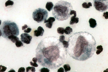
English: Color micrograph of the cytology of FIP-induced effusion. Magnification not specified; estimated to be 1000x.
Original caption: "The cytology of FIP effusion usually contains neutrophils, macrophages and lymphocytes." Image from "Feline Infectious Peritonitis: An Overview of Disease Transmission, Pathogenesis, Signs and Treatment With Emphasis on Diagnosis" ([1]) Clinical Pathology Clerkship Program |
| 日期 | 2005年9月30日 (原始上传日期) |
| 来源 | Transferred from en.wikipedia to Commons. |
| 作者 | 原上传者为英语维基百科的Bk0 |
许可协议
[编辑]

|
本文件的著作权人,允许任何人在适当地表明著作权人的姓名的前提下,以任何目的使用本文件。传播,演绎作品,商业用途及所有其他用途被允许。 |
|
|
原始上传日志
[编辑]
The original description page was here. All following user names refer to en.wikipedia.
- 2005-09-30 00:14 Bk0 350×234×8 (40687 bytes) Color micrograph of the cytology of [[Feline infectious peritonitis|FIP]]-induced effusion. Magnification not specified; estimated to be 1000x. Original caption: "The cytology of FIP effusion usually contains neutrophils, macrophages and lymphocytes." I
文件历史
点击某个日期/时间查看对应时刻的文件。
| 日期/时间 | 缩略图 | 大小 | 用户 | 备注 | |
|---|---|---|---|---|---|
| 当前 | 2007年12月29日 (六) 18:53 | | 350 × 234(40 KB) | Euthygenes(留言 | 贡献) | {{Information |Description={{en|Color micrograph of the cytology of FIP-induced effusion. Magnification not specified; estimated to be 1000x. Original caption: "The cytology of FIP effusion usually contains neutrophi |
您不可以覆盖此文件。
文件用途
没有页面使用本文件。
全域文件用途
以下其他wiki使用此文件:
- el.wikipedia.org上的用途
- en.wikipedia.org上的用途
- et.wikipedia.org上的用途
- fr.wikipedia.org上的用途
- hu.wikipedia.org上的用途
- ko.wikipedia.org上的用途
- tr.wikipedia.org上的用途
- zh.wikipedia.org上的用途