File:Gray491.png
Gray491.png (500 × 438 píxeles; tamaño de archivo: 63 kB; tipo MIME: image/png)
Leyendas
Leyendas
Resumen[editar]
| Descripción |
Deutsch: Blick von hinten auf das Herz. Darstellung von Henry Gray. |
||||||||||||||||||||
| Plate | 491 | ||||||||||||||||||||
| Fecha | antes de 1858 | ||||||||||||||||||||
| Fuente |
|
||||||||||||||||||||
| Autor |
|
||||||||||||||||||||
.jpg)
Libro[editar]
| Henry Gray: Gray's Anatomy
|
|||||||||||||||||||||||
|---|---|---|---|---|---|---|---|---|---|---|---|---|---|---|---|---|---|---|---|---|---|---|---|
| Autor |
|
-_Title_page.png) | |||||||||||||||||||||
| Director de edición |
Revised by Warren H. Lewis |
||||||||||||||||||||||
| Ilustrador |
|
||||||||||||||||||||||
| Título | |||||||||||||||||||||||
| Edición |
20 |
||||||||||||||||||||||
| Editorial | |||||||||||||||||||||||
| Object type |
edición, traducción o versión |
||||||||||||||||||||||
| Visión general | list of all the plates | ||||||||||||||||||||||
| Idioma |
inglés |
||||||||||||||||||||||
| Fecha de publicación |
1918 |
||||||||||||||||||||||
| Lugar de publicación |
Filadelfia / Nueva York |
||||||||||||||||||||||
| Fuente | Bartleby | ||||||||||||||||||||||

Licencia[editar]
Este archivo está en el dominio público porque es una exploración mecánica simple o fotocopia de un original en dominio público, o (con las pruebas disponibles) es tan similar a un documento escaneado o fotocopia que no se puede aplicar protección de derechos de autor. También puede suceder que los derechos de autor de esta imagen hayan expirado debido a la fecha de publicación o la muerte del autor (si es posible, añadirla aparte). El propio contenido original se encuentra en dominio público por las siguientes razones:
Esta etiqueta está diseñada para usarse cuando sea necesario afirmar que las mejoras (por ejemplo, brillo, contraste, juego de color, nitidez) no son de por sí suficientemente creativas para generar un nuevo derecho de autor. Se puede utilizar tanto cuando no se sabe si se han hecho mejoras como cuando las mejoras son claras, pero insuficientes. Para las imágenes primitivas sin contraste puede utilizar la plantilla {{PD-old}} adecuadamente insertada. Para utilizarla, véase Commons:Cuándo usar la etiqueta PD-scan.  | ||||
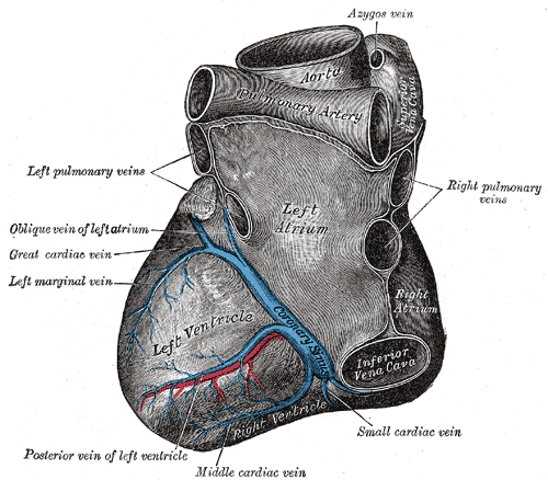
The coronary sinus is a collection of veins joined together to form a large vessel that collects blood from the myocardium of the heart. It is present in humans and other animals. It delivers deoxygenated blood to the Right atrium in conjunction with the superior and inferior vena cava.
The coronary sinus opens into the right atrium, between the inferior vena cava and the atrio-ventricular orifice. It returns the blood from the substance of the heart, and is protected by a semicircular fold of the lining membrane of the auricle, the coronary valve (the valve of Thebesius). The sinus, before entering the auricle, is considerably dilated - nearly to the size of the end of the little finger. Its wall is partly muscular, and at its junction with the great coronary vein is somewhat constricted and furnished with a valve consisting of two unequal segments.(Gray 462)
Location: It is located in the right atrium and runs transversely in the groove between the left atrium and ventricle on the posterior surface of the heart.
The coronary sinus orifice (opening) is just superior to the septal leaflet of the tricuspid valve. The coronary sinus orifice is also known as the ostium of the coronary sinus, and is guarded by the Thebesian valve.
Drainage: It receives blood mainly from the small, middle, great and oblique cardiac veins. It also receives blood from the left marginal vein and the left posterior ventricular vein. The anterior cardiac veins drain directly into the right atrium. (Some small veins drain into any of the four chambers of the heart.)
It drains into the right atrium on the posterior, inferior surface, medial to the inferior vena cava opening.
Historial del archivo
Haz clic sobre una fecha y hora para ver el archivo tal como apareció en ese momento.
| Fecha y hora | Miniatura | Dimensiones | Usuario | Comentario | |
|---|---|---|---|---|---|
| actual | 20:35 23 ene 2007 | | 500 × 438 (63 kB) | Pngbot (discusión | contribs.) | optimized with optipng |
| 06:26 11 feb 2006 |  | 500 × 438 (100 kB) | Arcadian (discusión | contribs.) | {{Gray's Anatomy plate}} |
No puedes sobrescribir este archivo.
Usos del archivo
Las siguientes páginas usan este archivo:
Uso global del archivo
Las wikis siguientes utilizan este archivo:
- Uso en ar.wikipedia.org
- Uso en bg.wikipedia.org
- Uso en bn.wikipedia.org
- Uso en bs.wikipedia.org
- Uso en cv.wikipedia.org
- Uso en de.wikibooks.org
- Uso en el.wikipedia.org
- Uso en en.wikipedia.org
- Coronary circulation
- Coronary sinus
- Oblique vein of the left atrium
- Posterior descending artery
- Circumflex branch of left coronary artery
- Vital heat
- Posterior interventricular sulcus
- Left marginal artery
- Smallest cardiac veins
- Vascular remodelling in the embryo
- Crux cordis
- User:Bob K31416/BH
- User:Walkerc84/sandbox
- User:Was a bee/Gray
- Uso en es.wikipedia.org
- Uso en fa.wikipedia.org
- Uso en it.wikipedia.org
- Uso en ja.wikipedia.org
- Uso en ko.wikipedia.org
- Uso en nl.wikipedia.org
- Uso en nn.wikipedia.org
- Uso en pl.wikipedia.org
Ver más uso global de este archivo.