File:Gray760.png
Gray760.png (500 × 323 Pixel, Dateigröße: 23 KB, MIME-Typ: image/png)
Bildtexte
Kurzbeschreibungen
Beschreibung[Bearbeiten]
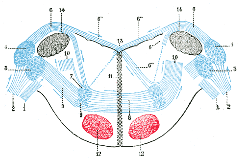
Terminal nuclei of the cochlear nerve, with their upper connections. (Schematic.) The vestibular nerve with its terminal nuclei and their efferent fibers have been suppressed. On the other hand, in order not to obscure the trapezoid body, the efferent fibers of the terminal nuclei on the right side have been resected in a considerable portion of their extent. The trapezoid body, therefore, shows only one-half of its fibers, viz., those which come from the left.
1. Vestibular nerve, divided at its entrance into the medulla oblongata.
2. Cochlear nerve.
3. Accessory nucleus of acoustic nerve.
4. Tuberculum acusticum.
5. Efferent fibers of accessory nucleus.
6. Efferent fibers of tuberculum acusticum, forming the striae medullares, with 6’, their direct bundle going to the superior olivary nucleus of the same side; 6’’, their decussating bundles going to the superior olivary nucleus of the opposite side.
7. Superior olivary nucleus.
8. Trapezoid body.
9. Trapezoid nucleus.
10. Central acoustic tract (lateral lemniscus).
11. Raphé.
12. Cerebrospinal fasciculus.
13. Fourth ventricle.
14. Inferior peduncle.
| Beschreibung | Terminal nuclei of the cochlear nerve, with their upper connections. (Schematic.) The vestibular nerve with its terminal nuclei and their efferent fibers have been suppressed. On the other hand, in order not to obscure the trapezoid body, the efferent fibers of the terminal nuclei on the right side have been resected in a considerable portion of their extent. The trapezoid body, therefore, shows only one-half of its fibers, viz., those which come from the left. 1. Vestibular nerve, divided at its entrance into the medulla oblongata. 2. Cochlear nerve. 3. Accessory nucleus of acoustic nerve. 4. Tuberculum acusticum. 5. Efferent fibers of accessory nucleus. 6. Efferent fibers of tuberculum acusticum, forming the striae medullares, with 6’, their direct bundle going to the superior olivary nucleus of the same side; 6’’, their decussating bundles going to the superior olivary nucleus of the opposite side. 7. Superior olivary nucleus. 8. Trapezoid body. 9. Trapezoid nucleus. 10. Central acoustic tract (lateral lemniscus). 11. Raphé. 12. Cerebrospinal fasciculus. 13. Fourth ventricle. 14. Inferior peduncle. (Testut.) | ||||||||||||||||||||
| Tafel | 760 | ||||||||||||||||||||
| Datum | vor 1858 | ||||||||||||||||||||
| Quelle |
|
||||||||||||||||||||
| Urheber |
|
||||||||||||||||||||
.jpg)
Buch[Bearbeiten]
| Henry Gray: Gray's Anatomy. - 20. Aufl. 1918
|
|||||||||||||||||||||||
|---|---|---|---|---|---|---|---|---|---|---|---|---|---|---|---|---|---|---|---|---|---|---|---|
| Urheber |
|
-_Title_page.png) | |||||||||||||||||||||
| Herausgeber |
Revised by Warren H. Lewis |
||||||||||||||||||||||
| Illustrator |
|
||||||||||||||||||||||
| Titel | |||||||||||||||||||||||
| Auflage |
20 |
||||||||||||||||||||||
| Verleger | |||||||||||||||||||||||
| Objektart |
Ausgabe oder Version |
||||||||||||||||||||||
| Seitenübersicht | list of all the plates | ||||||||||||||||||||||
| Sprache |
Englisch |
||||||||||||||||||||||
| Veröffentlichungsdatum |
1918 |
||||||||||||||||||||||
| Erscheinungsort |
Philadelphia / New York |
||||||||||||||||||||||
| Quelle | Bartleby | ||||||||||||||||||||||

Lizenz[Bearbeiten]
Dieses Bild ist gemeinfrei, weil es ein rein mechanischer Scan oder eine rein mechanische Fotografie eines gemeinfreien Originals ist, oder – wenn anders belegbar – es einem solchen Scan oder einer solchen Fotografie so ähnlich ist, dass das Entstehen eines Copyright-Schutzes nicht erwartet werden kann. Das Original selbst ist gemeinfrei aus folgendem Grund:
Dieser Hinweis wurde für Fälle entworfen, in denen erklärt werden muss, dass jegliche Verbesserungen (zum Beispiel von Helligkeit, Kontrast, Farbabgleich, Schärfe) für sich selbst nicht ausreichend sind, um ein neues Copyright zu schaffen. Er kann verwendet werden, wenn unbekannt ist, ob Verbesserungen vorgenommen wurden, oder wenn Verbesserungen erkennbar, aber nicht ausreichend sind. Für bekanntermaßen unbearbeitete Scans kannst Du statt dessen den passenden {{PD-old}}-Hinweis verwenden. Anwendungshinweise findest Du unter Commons:When to use the PD-scan tag.  | ||||
Dateiversionen
Klicke auf einen Zeitpunkt, um diese Version zu laden.
| Version vom | Vorschaubild | Maße | Benutzer | Kommentar | |
|---|---|---|---|---|---|
| aktuell | 20:52, 23. Jan. 2007 | | 500 × 323 (23 KB) | Pngbot (Diskussion | Beiträge) | optimized with optipng |
| 05:36, 29. Jan. 2006 |  | 500 × 323 (39 KB) | Arcadian (Diskussion | Beiträge) | {{Gray's Anatomy plate}} |
Du kannst diese Datei nicht überschreiben.
Dateiverwendung
Die folgenden 3 Seiten verwenden diese Datei:
Globale Dateiverwendung
Die nachfolgenden anderen Wikis verwenden diese Datei:
- Verwendung auf ar.wikipedia.org
- Verwendung auf bg.wikipedia.org
- Verwendung auf de.wikipedia.org
- Verwendung auf de.wikibooks.org
- Verwendung auf en.wikipedia.org
- Verwendung auf es.wikipedia.org
- Verwendung auf fa.wikipedia.org
- Verwendung auf ja.wikipedia.org
- Verwendung auf kk.wikipedia.org
- Verwendung auf nl.wikipedia.org
- Verwendung auf pl.wikipedia.org
- Verwendung auf zh.wikipedia.org