File:Gray881.png
Gray881.png (500 × 335 ピクセル、ファイルサイズ: 29キロバイト、MIME タイプ: image/png)
キャプション
キャプション
概要
[編集]| 解説 |
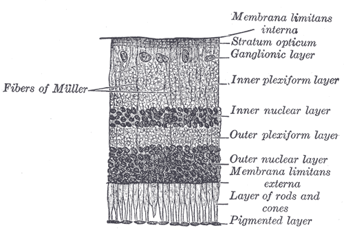
Original description from http://www.bartleby.com/107/225.html. The retina consists of an outer pigmented layer and an inner nervous stratum or retina proper. The pigmented layer consists of a single stratum of cells. When viewed from the outer surface these cells are smooth and hexagonal in shape; when seen in section each cell consists of an outer non-pigmented part containing a large oval nucleus and an inner pigmented portion which extends as a series of straight thread-like processes between the rods, this being especially the case when the eye is exposed to light. In the eyes of albinos the cells of this layer are destitute of pigment. Retina Proper —The nervous structures of the retina proper are supported by a series of nonnervous or sustentacular fibers, and, when examined microscopically by means of sections made perpendicularly to the surface of the retina, are found to consist of seven layers, named from within outward as follows:
|
||||||||||||||||||||
| Plate | 881 | ||||||||||||||||||||
| 日付 | 1858年より前 | ||||||||||||||||||||
| 原典 |
|
||||||||||||||||||||
| 作者 |
|
||||||||||||||||||||
| その他のバージョン | このファイルの派生的著作物: Gray881-ar.png | ||||||||||||||||||||
.jpg)
ブック
[編集]| ヘンリー・グレイ: グレイ解剖学 第20版
|
|||||||||||||||||||||||
|---|---|---|---|---|---|---|---|---|---|---|---|---|---|---|---|---|---|---|---|---|---|---|---|
| 作者 | -_Title_page.png) | ||||||||||||||||||||||
| 編者 |
Revised by Warren H. Lewis |
||||||||||||||||||||||
| 挿絵画家 |
|
||||||||||||||||||||||
| タイトル | |||||||||||||||||||||||
| 版 |
20 |
||||||||||||||||||||||
| 出版者 | |||||||||||||||||||||||
| 分野 |
バージョン、版、または翻訳 |
||||||||||||||||||||||
| ページ一覧 | list of all the plates | ||||||||||||||||||||||
| 言語 |
英語 |
||||||||||||||||||||||
| 出版日 |
1918年 |
||||||||||||||||||||||
| 出版地 |
フィラデルフィア / ニューヨーク |
||||||||||||||||||||||
| 原典 | Bartleby | ||||||||||||||||||||||

ライセンス
[編集]
この画像は、パブリックドメインにある原作品の機械的なスキャンか複写にすぎないこと、あるいは─入手可能な証拠から判断して─著作権保護が働かないような、機械的なスキャンまたは複写の結果と類似していることから、パブリックドメインにあります。原作品は下記の理由でパブリックドメインにあります。
使い方については、Commons:When to use the PD-scan tagをご覧ください。 注意:このタグはスキャンと複写にのみ適用されます。パブリックドメインの現物を一定の距離から写真撮影したものの場合は、{{PD-Art}}が適用できるかもしれません。詳しくはCommons:When to use the PD-scan tagをご覧ください。 | ||||
ファイルの履歴
過去の版のファイルを表示するには、その版の日時をクリックしてください。
| 日付と時刻 | サムネイル | 寸法 | 利用者 | コメント | |
|---|---|---|---|---|---|
| 現在の版 | 2007年1月23日 (火) 21:19 | | 500 × 335 (29キロバイト) | Pngbot (トーク | 投稿記録) | optimized with optipng |
| 2006年2月11日 (土) 03:14 |  | 500 × 335 (50キロバイト) | Arcadian (トーク | 投稿記録) | {{Gray's Anatomy plate}} |
このファイルは上書きできません。
ファイルの使用状況
以下の 3 ページがこのファイルを使用しています:
グローバルなファイル使用状況
以下に挙げる他のウィキがこの画像を使っています:
- ar.wikipedia.org での使用状況
- bg.wikipedia.org での使用状況
- bn.wikipedia.org での使用状況
- bs.wikipedia.org での使用状況
- Fotoosjetljive ganglijske ćelije
- Unutrašnja granična membrana
- Sloj nervnih vlakana
- Sloj ganglijskih ćelija
- Unutrašnji pleksiformni sloj
- Unutrašnji jezgarni sloj
- Vanjski pleksiformni sloj
- Vanjski jezgarni sloj
- Vanjska granična membrana
- Sloj štapića i čepića
- Pigmentni epitel mrežnjače
- Urođeno stacionarno noćno sljepilo
- ckb.wikipedia.org での使用状況
- de.wikibooks.org での使用状況
- en.wikipedia.org での使用状況
- Retina
- Neuropil
- Inner plexiform layer
- Layer of rods and cones
- Retinal pigment epithelium
- Inner nuclear layer
- External limiting membrane
- Internal limiting membrane
- Outer plexiform layer
- Outer nuclear layer
- Retinal nerve fiber layer
- Ganglion cell layer
- Congenital stationary night blindness
- Retinal regeneration
- User:Was a bee/Gray
- fa.wikipedia.org での使用状況
- he.wikipedia.org での使用状況
- hr.wikipedia.org での使用状況
- hy.wikipedia.org での使用状況
- it.wikipedia.org での使用状況
- ja.wikipedia.org での使用状況
- kn.wikipedia.org での使用状況
- ko.wikipedia.org での使用状況
このファイルのグローバル使用状況を表示する。