File:Hippel Lindau.gif
Μετάβαση στην πλοήγηση
Πήδηση στην αναζήτηση

Μέγεθος αυτής της προεπισκόπησης: 508 × 599 εικονοστοιχεία . Άλλες αναλύσεις: 204 × 240 εικονοστοιχεία | 407 × 480 εικονοστοιχεία | 805 × 949 εικονοστοιχεία.
Πρωτότυπο αρχείο (805 × 949 εικονοστοιχεία, μέγεθος αρχείου: 278 KB, τύπος MIME: image/gif)
Λεζάντες
Λεζάντες
Δεν ορίστηκε λεζάντα
Σύνοψη
[επεξεργασία]
| Περιγραφή |
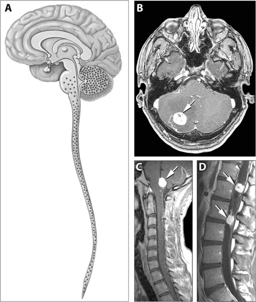
English: Distribution of Hemangioblastomas in the Central Nervous Systems of Study Patients
(A) Schematic representation of the distribution of CNS hemangioblastomas (red dots) in the 25 von Hippel-Lindau disease patients on MRI. Most (98%) of hemangioblastomas were found below the level of the tentorium in the cerebellum, brainstem, and spinal cord. (B–D) Contrast-enhanced MRI demonstrating representative locations of hemangioblastomas including the cerebellum (B), brainstem (C) and spinal cord (D). (B) Axial view through the cerebellum demonstrating a hyperintense enhancing hemangioblastoma (arrow) with surrounding edema (hypointense area surrounding the tumor) that frequently is associated with these lesions. (C) Sagittal view through the posterior fossa demonstrating a hyperintense enhancing brainstem (medullary) hemangioblastoma (arrow) with surrounding edema. (D) Sagittal view through the thoracic and lumbar spinal cord demonstrating two hyperintense enhancing hemangioblastomas (arrows). The superior tumor is associated with a large intraspinal cyst (syrinx) that is common with these neoplasms (arrowhead) |
| Πηγή | http://medicine.plosjournals.org/perlserv/?request=get-document&doi=10.1371/journal.pmed.0040060 |
| Δημιουργός |
Αδειοδότηση
[επεξεργασία]
|
Το αρχείο διανέμεται υπό την άδεια Creative Commons Αναφορά προέλευσης 2.5 Γενική
|
This file was published in a Public Library of Science journal. Their website states that the content of all PLOS journals is published under the Creative Commons Attribution 4.0 license (or its previous version depending on the publication date), unless indicated otherwise.
|
Ιστορικό αρχείου
Πατήστε σε μια ημερομηνία/ώρα για να δείτε το αρχείο όπως εμφανιζόταν εκείνη την χρονική στιγμή.
| Ημερομηνία/Ώρα | Μικρογραφία | Διαστάσεις | Χρήστης | Σχόλιο | |
|---|---|---|---|---|---|
| τρέχον | 13:43, 31 Μαΐου 2007 | | 805 × 949 (278 KB) | Filip em (συζήτηση | Συνεισφορά) | Distribution of Hemangioblastomas in the Central Nervous Systems of Study Patients (A) Schematic representation of the distribution of CNS hemangioblastomas (red dots) in the 25 von Hippel-Lindau disease patients on MRI. Most (98%) of hemangioblastomas w |
Δεν μπορείτε να αντικαταστήσετε αυτό το αρχείο.
Χρήση αρχείου
Δεν υπάρχουν σελίδες που χρησιμοποιούν αυτό το αρχείο.
Καθολική χρήση αρχείου
Τα ακόλουθα άλλα wiki χρησιμοποιούν αυτό το αρχείο:
- Χρήση σε ar.wikipedia.org
- Χρήση σε bs.wikipedia.org
- Χρήση σε ca.wikipedia.org
- Χρήση σε de.wikipedia.org
- Χρήση σε de.wikibooks.org
- Χρήση σε el.wikipedia.org
- Χρήση σε en.wikipedia.org
- Χρήση σε hy.wikipedia.org
- Χρήση σε it.wikipedia.org
- Χρήση σε ja.wikipedia.org
- Χρήση σε ru.wikipedia.org
- Χρήση σε sk.wikipedia.org
- Χρήση σε uz.wikipedia.org
- Χρήση σε zh.wikipedia.org