File:Mitochondria, mammalian lung - TEM.jpg
Aller à la navigation
Aller à la recherche
Pas de plus haute résolution disponible.
Mitochondria,_mammalian_lung_-_TEM.jpg (640 × 480 pixels, taille du fichier : 96 kio, type MIME : image/jpeg)
Légendes
Légendes
Ajoutez en une ligne la description de ce que représente ce fichier
Description[modifier]
| Description |
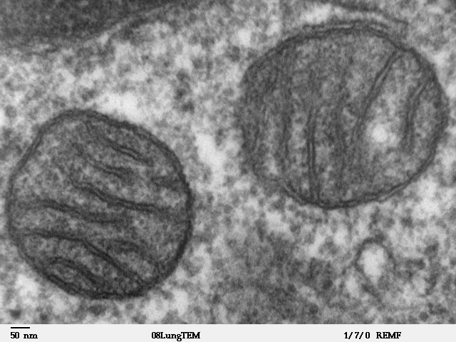
Transmission electron microscope image of a thin section cut through an area of mammalian lung tissue. The high magnification image shows two mitochondria. JEOL 100CX TEM |
| Source | |
| Auteur | Louisa Howard |
| Autorisation (Réutilisation de ce fichier) |
PD |
Conditions d’utilisation[modifier]
| Cette œuvre a été placée dans le domaine public par son auteur, Louisa Howard. Ceci s’applique dans le monde entier. Dans certains pays, ceci peut ne pas être possible ; dans ce cas : Louisa Howard accorde à toute personne le droit d’utiliser cette œuvre dans n’importe quel but, sans aucune condition, sauf celles requises par la loi.
|
Historique du fichier
Cliquer sur une date et heure pour voir le fichier tel qu'il était à ce moment-là.
| Date et heure | Vignette | Dimensions | Utilisateur | Commentaire | |
|---|---|---|---|---|---|
| actuel | 16 mai 2008 à 15:09 | | 640 × 480 (96 kio) | Vojtěch Dostál (d | contributions) | Reverted to version as of 15:37, 5 October 2006, my fault |
| 16 mai 2008 à 12:51 |  | 640 × 433 (84 kio) | Vojtěch Dostál (d | contributions) | {{Information |Description=Transmission electron microscope image of a thin section cut through an area of mammalian lung tissue. The high magnification image shows a mitochondria. JEOL 100CX TEM |Source= * http://remf.dartmouth.edu/imagesindex.html * h | |
| 16 mai 2008 à 12:47 |  | 640 × 453 (86 kio) | Vojtěch Dostál (d | contributions) | {{Information |Description=Transmission electron microscope image of a thin section cut through an area of mammalian lung tissue. The high magnification image shows a mitochondria. JEOL 100CX TEM |Source= * http://remf.dartmouth.edu/imagesindex.html * h | |
| 5 octobre 2006 à 15:37 |  | 640 × 480 (96 kio) | Patho (d | contributions) | {{Information |Description=Transmission electron microscope image of a thin section cut through an area of mammalian lung tissue. The high magnification image shows a mitochondria. JEOL 100CX TEM |Source= * http://remf.dartmouth.edu/imagesindex.html * h |
Vous ne pouvez pas remplacer ce fichier.
Utilisations locales du fichier
Les 4 pages suivantes utilisent ce fichier :
.jpg)
Utilisations du fichier sur d’autres wikis
Les autres wikis suivants utilisent ce fichier :
- Utilisation sur ar.wikipedia.org
- Utilisation sur az.wiktionary.org
- Utilisation sur be.wikipedia.org
- Utilisation sur bg.wikipedia.org
- Utilisation sur bn.wikipedia.org
- Utilisation sur br.wikipedia.org
- Utilisation sur bs.wikipedia.org
- Utilisation sur ca.wikipedia.org
- Utilisation sur cdo.wikipedia.org
- Utilisation sur da.wikipedia.org
- Utilisation sur de.wikibooks.org
- Utilisation sur el.wikipedia.org
- Utilisation sur en.wikipedia.org
- Utilisation sur en.wikibooks.org
- Utilisation sur en.wikiversity.org
- User:Jtwsaddress42/Projects/Project 1
- User:Jtwsaddress42/Projects/Project 1/Parts
- User:Jtwsaddress42/Projects/Project 1/Parts/Part 3
- User:Jtwsaddress42/Projects/Project 1/Chapters/Chapter 10
- User:Jtwsaddress42/Projects/Project 1/Sections/Chapter 10/Phase II - The Oxygen Crisis and the Rise of the Aerobic Bioshphere (1.9-0.95 bya)
- User:Jtwsaddress42/Clade
- User:Jtwsaddress42/Clade/Gracilicutes to Proteobacteria
- Utilisation sur en.wiktionary.org
- Utilisation sur es.wikipedia.org
- Utilisation sur et.wikipedia.org
- Utilisation sur eu.wikipedia.org
- Utilisation sur ext.wikipedia.org
- Utilisation sur fa.wikipedia.org
- Utilisation sur fr.wikipedia.org
Voir davantage sur l’utilisation globale de ce fichier.