File:Mitochondria, mammalian lung - TEM.jpg
跳至導覽
跳至搜尋
無更高解析度可提供。
Mitochondria,_mammalian_lung_-_TEM.jpg (640 × 480 像素,檔案大小:96 KB,MIME 類型:image/jpeg)
說明
說明
添加單行說明來描述出檔案所代表的內容
摘要
[編輯]
| 描述 |
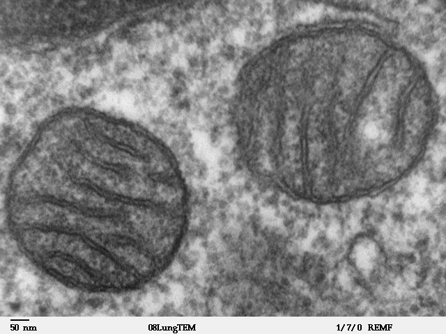
Transmission electron microscope image of a thin section cut through an area of mammalian lung tissue. The high magnification image shows two mitochondria. JEOL 100CX TEM |
| 來源 | |
| 作者 | Louisa Howard |
| 授權許可 (重用此檔案) |
PD |
授權條款
[編輯]
| 此作品已由其作者,Louisa Howard,釋出至公有領域。此授權條款在全世界均適用。 這可能在某些國家不合法,如果是的話: Louisa Howard授予任何人有權利使用此作品於任何用途,除受法律約束外,不受任何限制。
|
檔案歷史
點選日期/時間以檢視該時間的檔案版本。
| 日期/時間 | 縮圖 | 尺寸 | 使用者 | 備註 | |
|---|---|---|---|---|---|
| 目前 | 2008年5月16日 (五) 15:09 | | 640 × 480(96 KB) | Vojtěch Dostál(留言 | 貢獻) | Reverted to version as of 15:37, 5 October 2006, my fault |
| 2008年5月16日 (五) 12:51 |  | 640 × 433(84 KB) | Vojtěch Dostál(留言 | 貢獻) | {{Information |Description=Transmission electron microscope image of a thin section cut through an area of mammalian lung tissue. The high magnification image shows a mitochondria. JEOL 100CX TEM |Source= * http://remf.dartmouth.edu/imagesindex.html * h | |
| 2008年5月16日 (五) 12:47 |  | 640 × 453(86 KB) | Vojtěch Dostál(留言 | 貢獻) | {{Information |Description=Transmission electron microscope image of a thin section cut through an area of mammalian lung tissue. The high magnification image shows a mitochondria. JEOL 100CX TEM |Source= * http://remf.dartmouth.edu/imagesindex.html * h | |
| 2006年10月5日 (四) 15:37 |  | 640 × 480(96 KB) | Patho(留言 | 貢獻) | {{Information |Description=Transmission electron microscope image of a thin section cut through an area of mammalian lung tissue. The high magnification image shows a mitochondria. JEOL 100CX TEM |Source= * http://remf.dartmouth.edu/imagesindex.html * h |
無法覆蓋此檔案。
檔案用途
下列4個頁面有用到此檔案:
.jpg)
全域檔案使用狀況
以下其他 wiki 使用了這個檔案:
- ar.wikipedia.org 的使用狀況
- az.wiktionary.org 的使用狀況
- be.wikipedia.org 的使用狀況
- bg.wikipedia.org 的使用狀況
- bn.wikipedia.org 的使用狀況
- br.wikipedia.org 的使用狀況
- bs.wikipedia.org 的使用狀況
- ca.wikipedia.org 的使用狀況
- cdo.wikipedia.org 的使用狀況
- da.wikipedia.org 的使用狀況
- de.wikibooks.org 的使用狀況
- el.wikipedia.org 的使用狀況
- en.wikipedia.org 的使用狀況
- en.wikibooks.org 的使用狀況
- en.wikiversity.org 的使用狀況
- User:Jtwsaddress42/Projects/Project 1
- User:Jtwsaddress42/Projects/Project 1/Parts
- User:Jtwsaddress42/Projects/Project 1/Parts/Part 3
- User:Jtwsaddress42/Projects/Project 1/Chapters/Chapter 10
- User:Jtwsaddress42/Projects/Project 1/Sections/Chapter 10/Phase II - The Oxygen Crisis and the Rise of the Aerobic Bioshphere (1.9-0.95 bya)
- User:Jtwsaddress42/Clade
- User:Jtwsaddress42/Clade/Gracilicutes to Proteobacteria
- en.wiktionary.org 的使用狀況
- es.wikipedia.org 的使用狀況
- et.wikipedia.org 的使用狀況
- eu.wikipedia.org 的使用狀況
- ext.wikipedia.org 的使用狀況
檢視此檔案的更多全域使用狀況。