File:Nucleosome core particle 1EQZ v.5.jpg
Nucleosome_core_particle_1EQZ_v.5.jpg (620 × 560 εικονοστοιχεία, μέγεθος αρχείου: 99 KB, τύπος MIME: image/jpeg)
Λεζάντες
Λεζάντες
Σύνοψη
[επεξεργασία]
| Περιγραφή |
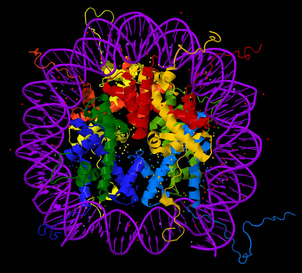
English: Nucleosome core particle, crystal structure (PDB ID: 1EQZ). Histones H2A, H2B, H3 and H4 are coloured. This image was created with Jmol (an open-source Java viewer for chemical structures in 3D http://www.jmol.org/) and Adobe Photoshop Elements. |
| Ημερομηνία | |
| Πηγή |
Own work using: the Protein Data Bank (PDB) structural data: |
| Δημιουργός | Darekk2 using the cited above Protein Data Bank (PDB) structural data |
Αδειοδότηση
[επεξεργασία]
Attribution: The author of the work, the original authors of the Protein Data Bank (PDB) structural data and the molecular graphics program used. PDB data and software citation example:
Image of PDB (Protein Data Bank) ID 1EQZ (Harp, J.M., Hanson, B.L., Timm, D.E., Bunick, G.J. (2000) Asymmetries in the nucleosome core particle at 2.5 A resolution. Acta Crystallogr., Sect.D 56: 1513-1534) created with Jmol (an open-source Java viewer for chemical structures in 3D. http://www.jmol.org/).
The RCSB Protein Data Bank (PDB) states on its website in the Policies & References section:
http://www.rcsb.org/pdb/static.do?p=general_information/about_pdb/policies_references.html archive copy at the Wayback Machine
Data files contained in the PDB archive (ftp://ftp.wwpdb.org) are free of all copyright restrictions and made fully and freely available for both non-commercial and commercial use. Users of the data should attribute the original authors of that structural data. (...) Images created using PDB data and other software should cite the PDB ID and the molecular graphics program used.
the original authors of the Protein Data Bank (PDB) structural data
and the molecular graphics program used
- Είστε ελεύθερος:
- να μοιραστείτε – να αντιγράψετε, διανέμετε και να μεταδώσετε το έργο
- να διασκευάσετε – να τροποποιήσετε το έργο
- Υπό τις ακόλουθες προϋποθέσεις:
- αναφορά προέλευσης – Θα πρέπει να κάνετε κατάλληλη αναφορά, να παρέχετε σύνδεσμο για την άδεια και να επισημάνετε εάν έγιναν αλλαγές. Μπορείτε να το κάνετε με οποιοδήποτε αιτιολογήσιμο λόγο, χωρίς όμως να εννοείται με οποιονδήποτε τρόπο ότι εγκρίνουν εσάς ή τη χρήση του έργου από εσάς.
- παρόμοια διανομή – Εάν αλλάξετε, τροποποιήσετε ή δημιουργήσετε πάνω στο έργο αυτό, μπορείτε να διανείμετε αυτό που θα προκύψει μόνο υπό τους όρους της ίδιας ή συμβατής άδειας με το πρωτότυπο.
Ιστορικό αρχείου
Πατήστε σε μια ημερομηνία/ώρα για να δείτε το αρχείο όπως εμφανιζόταν εκείνη την χρονική στιγμή.
| Ημερομηνία/Ώρα | Μικρογραφία | Διαστάσεις | Χρήστης | Σχόλιο | |
|---|---|---|---|---|---|
| τρέχον | 22:16, 9 Οκτωβρίου 2012 | | 620 × 560 (99 KB) | Darekk2 (συζήτηση | Συνεισφορά) | changed colors and rotated 180 deg |
| 12:25, 9 Οκτωβρίου 2012 |  | 596 × 567 (94 KB) | Darekk2 (συζήτηση | Συνεισφορά) | User created page with UploadWizard |
Δεν μπορείτε να αντικαταστήσετε αυτό το αρχείο.
Χρήση αρχείου
Η ακόλουθη σελίδα χρησιμοποιεί προς αυτό το αρχείο:
Καθολική χρήση αρχείου
Τα ακόλουθα άλλα wiki χρησιμοποιούν αυτό το αρχείο:
- Χρήση σε ar.wikipedia.org
- Χρήση σε bs.wikipedia.org
- Χρήση σε ca.wikipedia.org
- Χρήση σε el.wikipedia.org
- Χρήση σε en.wikipedia.org
- Χρήση σε es.wikipedia.org
- Χρήση σε ja.wikipedia.org
- Χρήση σε vi.wikipedia.org