File:Oncocytoma of the Salivary Gland.jpg
Oncocytoma_of_the_Salivary_Gland.jpg (550 × 335 Pixel, Dateigröße: 25 KB, MIME-Typ: image/jpeg)
Bildtexte
Kurzbeschreibungen
Beschreibung
[Bearbeiten]
| Beschreibung |
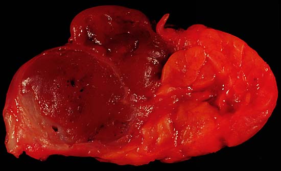
Oncocytoma of the Salivary Gland This lesion presented as a lateral anterior neck mass. At surgery, it was found to be a soft 3.0 x 2.1 x 1.8 cm tumor of the submandibular salivary gland. The photo shows the characteristic dark color of an oncocytoma, a rare type of benign neoplasm, at the left side of the image (the normal lobulated salivary gland tissue is to the right). Excision is curative. Since this specimen was photographed in the fresh state, it is various shades of red due to blood staining. A little formalin fixation would be expected to better emphasize the color difference between the tumor and the normal gland tissue. The photo was shot with a Minolta X-370 with 100mm Rokkor bellows lens on Kodak Elite ISO 100 daylight-balanced transparency film. I used a blue filter to compensate for the tungsten illumination. Photograph by Ed Uthman, MD. Public domain. Posted 19 May 00 |
| Quelle | http://web2.airmail.net/uthman/specimens/index.html |
| Urheber | |
| Genehmigung (Weiternutzung dieser Datei) |
PD |
Lizenz
[Bearbeiten]
| Dieses Werk wurde von seinem Urheber Ed Uthman als gemeinfrei veröffentlicht. Dies gilt weltweit. In manchen Staaten könnte dies rechtlich nicht möglich sein. Sofern dies der Fall ist: Ed Uthman gewährt jedem das bedingungslose Recht, dieses Werk für jedweden Zweck zu nutzen, es sei denn, Bedingungen sind gesetzlich erforderlich.
|
Dateiversionen
Klicke auf einen Zeitpunkt, um diese Version zu laden.
| Version vom | Vorschaubild | Maße | Benutzer | Kommentar | |
|---|---|---|---|---|---|
| aktuell | 09:53, 5. Jun. 2006 | | 550 × 335 (25 KB) | Patho (Diskussion | Beiträge) | {{Information| |Description=Oncocytoma of the Salivary Gland This lesion presented as a lateral anterior neck mass. At surgery, it was found to be a soft 3.0 x 2.1 x 1.8 cm tumor of the submandibular salivary gland. The photo shows the characteristic dar |
Du kannst diese Datei nicht überschreiben.
Dateiverwendung
Keine Seiten verwenden diese Datei.
Globale Dateiverwendung
Die nachfolgenden anderen Wikis verwenden diese Datei:
- Verwendung auf de.wikipedia.org
- Verwendung auf de.wikibooks.org
- Verwendung auf en.wikipedia.org
- Verwendung auf fr.wikipedia.org
- Verwendung auf pl.wikipedia.org