File:Paramecium.jpg
Pereiti į navigaciją
Jump to search

Šios peržiūros dydis: 610 × 599 taškų. Kitos 3 rezoliucijos: 244 × 240 taškų | 488 × 480 taškų | 751 × 738 taškų.
Pradinė rinkmena (751 × 738 taškų, rinkmenos dydis: 186 KiB, MIME tipas: image/jpeg)
Captions
Captions
Add a one-line explanation of what this file represents
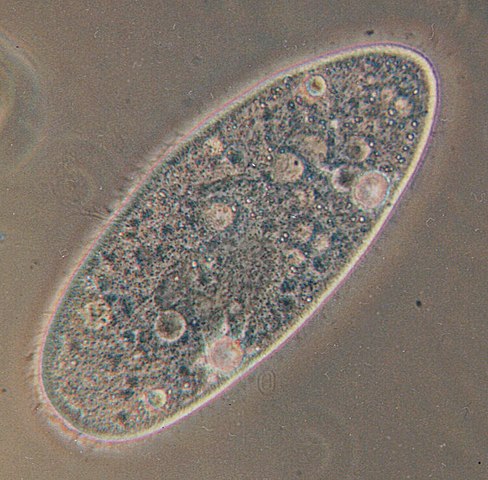
A Paramecium protist.
Aprašymas[keisti]
| Aprašymas |
Deutsch: Paramecium aurelia - Optisches Mikroskop. Paramecium aurelia, der bekannteste von allen ciliaten. Die Blasen innerhalb der Zelle sind Vakuolen. Die gesamte Oberfläche ist mit Wimpern umgeben, die durch ihre schnelle Bewegung verwischt werden.
English: Paramecium aurelia. Optical microscope. Paramecium aurelia, the best known of all ciliates. The bubbles throughout the cell are vacuoles. The entire surface is covered in cilia, which are blurred by their rapid movement.
Français : Paramecium aurelia. Microscope optique. Le plus connu des ciliés. Les bulles que vous voyez sont des vacuoles. Tout le corps est couvert par des cils, qui sont flous sur l'image à cause de leurs mouvements rapides.
Polski: Paramecium aurelia - pantofelek, najbardziej znany ze wszystkich orzęsków. Bąbelki w środku komórki to wodniczki. Cała powierzchnia pantofelka pokryta jest rzęskami, które są na fotografii zamazane ze względu na ich szybki ruch.
Српски / srpski: Paramecium aurelia, najpoznatiji od svih trepljara pod optičkim mikroskopom. "Mehurići" u ćeliji paramecijuma su vakuole. Cela površina tela je prekrivena trepljama, koje su na slici mutne zbog toga što se brzo pokreću.
Türkçe: Paramecium aurelia - optik mikroskop. Paramecium aurelia, tüm siliyalılar içinde en çok bilinen türdür. Hücre boyunca yuvarlak olarak izlenen oluşumlar, vakuollerdir. Hücrenin tüm yüzeyi, hızlı hareketlerinden dolayı bulanık görüntü vermiş olan siliya ile kaplıdır. |
| Data | |
| Šaltinis | Originally uploaded to the English Wikipedia, where it was made by Barfooz. |
| Autorius | Barfooz at the English Wikipedia. |
| Kitos versijos | Transparent |
Licencija[keisti]

|
Suteikiamas leidimas kopijuoti, platinti ir/ar redaguoti šį dokumentą pagal GNU Free Documentation licencijos versijos 1.2 ar bet kurios vėlesnės versijos sąlygas, publikuotas Free Software Foundation; be nekintamų dalių, be priekinių ir galinių tekstinių žymų viršeliuose. Šios licencijos kopija įtraukta dalyje, pavadintoje GNU Free Documentation License. |
| Šiam failui taikoma Creative Commons Attribution-Share Alike 3.0 Unported licencija. | ||
| ||
| Licencijos šablonas buvo priskirtas šiam failui kaip GFDL licencijos atnaujinimo dalis. |
Soft scrubbed view
Originalus įkėlimo įrašas[keisti]
Originally uploaded to English Wikipedia.
- 23:11, 27 October 2004 . . Barfooz (Talk) . . 751x738 (190517 bytes) (Paramecium viewed under a microscope)
- 15:19, 28 June 2004 . . Josh Grosse (Talk) . . 236x152 (3913 bytes) (Reverted to earlier revision)
- 15:19, 28 June 2004 . . Josh Grosse (Talk) . . 236x152 (5129 bytes) (Reverted to earlier revision)
- 15:13, 28 June 2004 . . Josh Grosse (Talk) . . 236x152 (3913 bytes) (Better image, created by self)
- 20:04, 10 October 2003 . . Josh Grosse (Talk) . . 236x152 (5129 bytes)
Rinkmenos istorija
Paspauskite ant datos/laiko, kad pamatytumėte rinkmeną tokią, kokia ji buvo tuo metu.
| Data/Laikas | Miniatiūra | Matmenys | Naudotojas | Paaiškinimas | |
|---|---|---|---|---|---|
| dabartinis | 20:46, 31 gegužės 2005 | | 751 × 738 (186 KiB) | Luis Fernández García (aptarimas | indėlis) | ''Paramecium aurelia''. Optical microscope Source: English Wikipedia (http://en.wikipedia.org/wiki/Image:Paramecium.jpg) |
Jūs negalite perrašyti šios rinkmenos.
Rinkmenos naudojimas
Šie puslapiai naudoja šią rinkmeną:
Visuotinis rinkmenos naudojimas
Ši rinkmena naudojama šiose viki svetainėse:
- Naudojama als.wikipedia.org
- Naudojama an.wikipedia.org
- Naudojama ar.wikipedia.org
- خلية
- طلائعيات
- بوابة:علم الأحياء الخلوي والجزيئي
- بوابة:علم الأحياء الخلوي والجزيئي/مواضيع علم الأحياء الخلوي والجزيئي
- ويكيبيديا:قوالب/قوالب المعلومات/علوم
- براميسيوم
- خصائص الكائنات الحية
- دوارات
- مكورة عنقودية ذهبية
- ضمة الكوليرا
- مملكة (تصنيف)
- مكورة دقيقة
- قالب:بذرة أحياء دقيقة
- عصوية رقيقة
- مبيضة بيضاء
- غيري التغذية
- لولبية شاحبة
- مكورات عنقودية
- أوليغيلا
- مفطورة
- كمون الفيروس
- فيروس موجه للعصب
- بكتيريا زرقاء
- محلل (أحياء)
- عقدية
- كوكسيلة بورنيتية
- مستحرة مائية
- متسلسلة (بكتيريا)
- شعاوات
- متسلسلات (بكتيريا)
- بكتيريا زرنيخية
- تلوين تسيل-نلسن
- أغار مغذي
- أغار مولر-هينتون
- اختبار السكريات الثلاثية والحديد
- أغار ستريميد
- أغار البطاطس بالدكستروز
- علم الأحياء الدقيقة الطبي
- الجينايت
- موضع الشق (بكتيريا)
- إقصاء تنافسي
- بروتين نووي
- هيكسون
- بوليميراز الرنا المعتمدة على الرنا
- بروتين سكري 120
- حمض نووي ريبوزي ناقل
- بكتيريا هوائية إجبارية
- تخطيط الاستنماء
Žiūrėti visuotinį šios rinkmenos naudojimą.