File:Parasite170144-fig16 Spermatozoon ultrastructure in Digenea, Monopisthocotylea, Polyopisthocotylea.png
Salta a la navegació
Salta a la cerca
Mida d'aquesta previsualització: 800 × 358 píxels. Altres resolucions: 320 × 143 píxels | 640 × 286 píxels | 1.024 × 458 píxels | 1.280 × 572 píxels | 2.560 × 1.145 píxels | 6.274 × 2.806 píxels.
Fitxer original (6.274 × 2.806 píxels, mida del fitxer: 757 Ko, tipus MIME: image/png)
Llegendes
Llegendes
Afegeix una explicació d'una línia del que representa aquest fitxer
Resum
[modifica]
| Descripció |
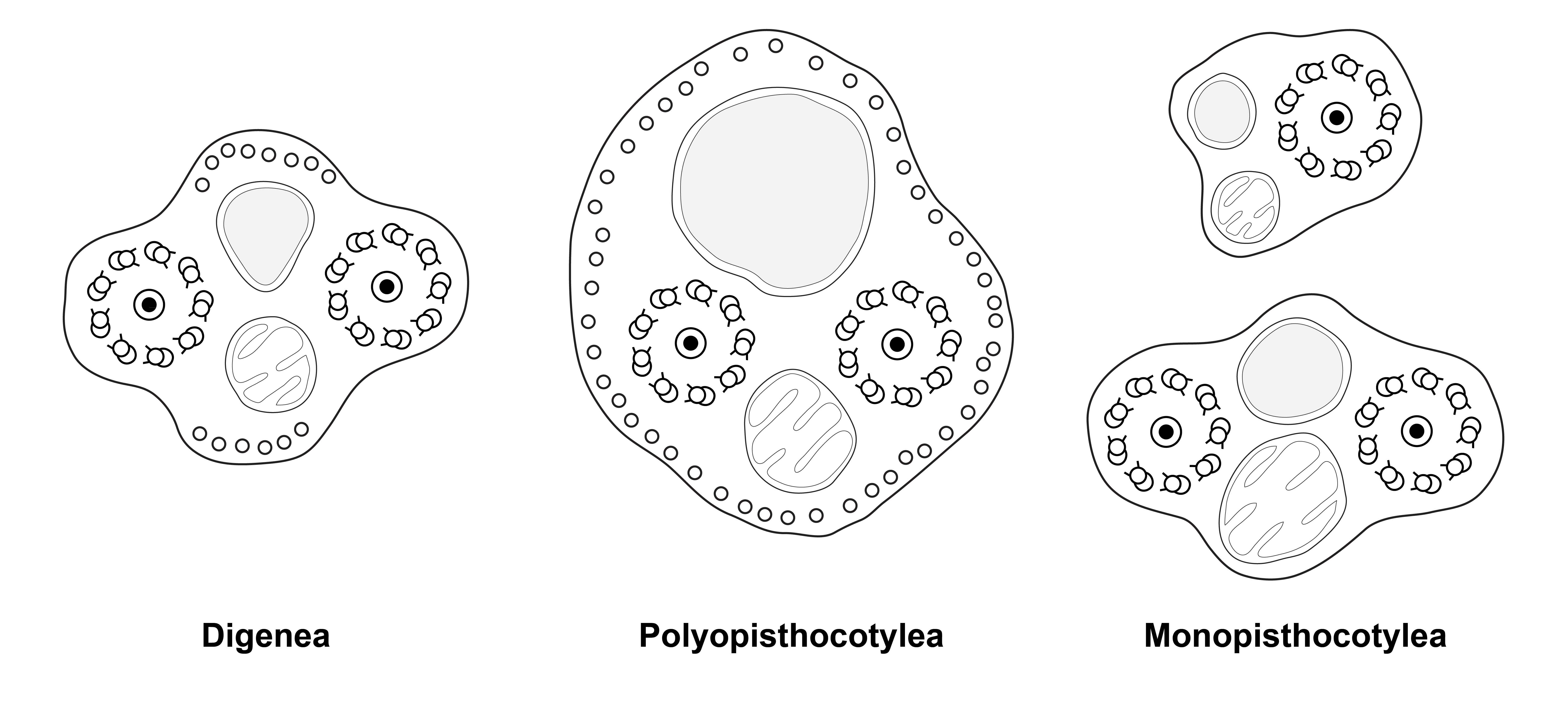
English: Figure 16 of the paper. Diagrams of spermatozoa (redrawn from Justine, 1991). Caption adapted from original caption of figure. Diagrams were drawn from original micrographs of transverse sections. Digenea. Dorsal and ventral microtubules are present (proposed as a synapomorphy for the Cercomeridea). There are no lateral microtubules (symplesiomorphic compared with the synapomorphy for the polyopisthocotylean Monogenea). Polyopisthocotylea. Dorsal and ventral microtubules are present (synapomorphy for Cercomeridea). Note the presence of lateral microtubules (proposed as a synapomorphy for the Polyopisthocotylea). Monopisthocotylea (uniflagellate and biflagellate). Microtubules are absent from the principal region of the spermatozoon, which is interpreted as (i) the absence of dorsal and ventral microtubules, a reversal of the synapomorphy for the Cercomeridea; and (ii) the absence of lateral microtubules, the symplesiomorphic state versus the synapomorphy for the Polyopisthocotylea. |
| Data | |
| Font | (2018). "Spermiogenesis and spermatozoon ultrastructure in basal polyopisthocotylean monogeneans, Hexabothriidae and Chimaericolidae, and their significance for the phylogeny of the Monogenea". Parasite 25: 7. DOI:10.1051/parasite/2018007. ISSN 1776-1042. |
| Autor | Jean-Lou Justine and Larisa G. Poddubnaya |
Llicència
[modifica]
This file is licensed under the Creative Commons Attribution 4.0 International license.
|
 This file was published in the scientific journal Parasite. Their website states that all content of the journal including and after 2013 is published under the Creative Commons Attribution 4.0 license.
|
Historial del fitxer
Cliqueu una data/hora per veure el fitxer tal com era aleshores.
| Data/hora | Miniatura | Dimensions | Usuari/a | Comentari | |
|---|---|---|---|---|---|
| actual | 16:58, 15 feb 2018 | | 6.274 × 2.806 (757 Ko) | Jeanloujustine (discussió | contribucions) | User created page with UploadWizard |
No podeu sobreescriure aquest fitxer.
Ús del fitxer
La pàgina següent utilitza aquest fitxer:
Ús global del fitxer
Utilització d'aquest fitxer en altres wikis:
- Utilització a ca.wikipedia.org
- Utilització a en.wikipedia.org
- Utilització a fa.wikipedia.org
- Utilització a tr.wikipedia.org
- Utilització a www.wikidata.org