File:Rostral migratory stream mouse.jpg
Aller à la navigation
Aller à la recherche
Taille de cet aperçu : 347 × 599 pixels. Autres résolutions : 139 × 240 pixels | 278 × 480 pixels | 445 × 768 pixels | 1 196 × 2 063 pixels.
Fichier d’origine (1 196 × 2 063 pixels, taille du fichier : 241 kio, type MIME : image/jpeg)
Légendes
Légendes
Ajoutez en une ligne la description de ce que représente ce fichier
| Description |
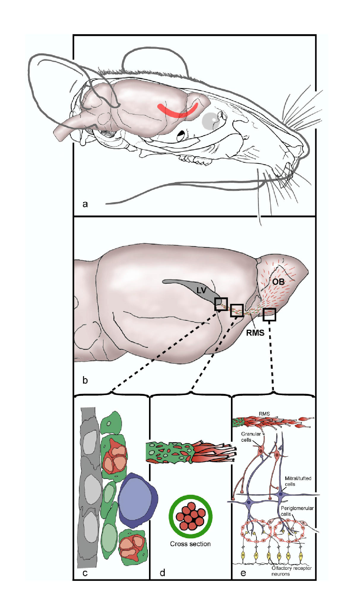
English: (a) Head of a mouse showing the location of the brain and the rostral migratory stream, RMS, (in red) along which newly generated neuroblasts migrate from the SVZ of the lateral ventricle into the olfactory bulb (OB). (b) The migration of newly generated neuroblasts begins at the lateral ventricle, continues along the RMS and terminated in the OB, where mature interneuron populations are generated. (c) Schematic based on electron microscopy showing the cytoarchitecture of the SVZ along the ventricle. Ependymal cells (gray) form a monolayer along the ventricle with astrocytes (green), neuroblasts (red) and transitory amplifying progenitors (TAP) (purple) comprising the SVZ. (d) Schematic showing the migration of neuroblasts along the RMS. Astrocytes (green) ensheath the migrating neuroblasts (red) and are thought to restrict and contain the neuroblasts to their specific pathway. (e) Migrating neuroblasts enter the OB, migrate radially and give rise to granule or periglomerular cells.
Русский: (A): Голова мыши. Показано расположение рострального миграционного тракта в мозге (красная полоса). По этому пути мигрируют свежесозданные нейробласты из субвентрикулярной зоны в обонятельную луковицу. (B): Миграция новых нейробластов начинается с бокового желудочка, затем клетки проходят весь РМТ до обонятельной луковицы, в которой генерируются взрослые популяции нейронов. (C): Схема, основанная на данных электронной микроскопии, демонстрирующая цироархитектуру субвентрикулярной зоны вдоль желудочка. Эпендимоциты (серые) формируют моно-слой а астроциты (зел), нейробласты (красн) и транзиторные делящиеся предшественники (TAP, пурпурные), составляют тело субвентрикулярной зоны. (D): Схема нейромиграции в тракте. Астроциты (зел.) окутывают мигрирующие нейробласты (красн.) и, как считается, ограничивают их свободу, направляя по строго предопределенному пути. (E): Мигрирующие нейробласты достигли обон. луковицы, теперь они мигрируют радиально и закрепляются, превращаясь в гранульные или перигломерулярные клетки. |
| Date | |
| Source | Lennington et al. Neural stem cells and the regulation of adult neurogenesis. Reproductive Biology and Endocrinology 2003 1:99 doi:10.1186/1477-7827-1-99 |
| Auteur | Jessica B Lennington, Zhengang Yang and Joanne C Conover |
| Autorisation (Réutilisation de ce fichier) |
© 2003 Lennington et al; licensee BioMed Central Ltd. This is an Open Access article: verbatim copying and redistribution of this article are permitted in all media for any purpose, provided this notice is preserved along with the article's original URL. |
| Autres versions | Œuvres dérivées de ce fichier : Rostral migratory stream mouse cropped.jpg |
Ce fichier est disponible selon les termes de la licence Creative Commons Attribution 2.0 Générique.
- Vous êtes libre :
- de partager – de copier, distribuer et transmettre cette œuvre
- d’adapter – de modifier cette œuvre
- Sous les conditions suivantes :
- paternité – Vous devez donner les informations appropriées concernant l'auteur, fournir un lien vers la licence et indiquer si des modifications ont été faites. Vous pouvez faire cela par tout moyen raisonnable, mais en aucune façon suggérant que l’auteur vous soutient ou approuve l’utilisation que vous en faites.
Historique du fichier
Cliquer sur une date et heure pour voir le fichier tel qu'il était à ce moment-là.
| Date et heure | Vignette | Dimensions | Utilisateur | Commentaire | |
|---|---|---|---|---|---|
| actuel | 4 août 2008 à 11:41 | | 1 196 × 2 063 (241 kio) | CopperKettle (d | contributions) | {{Information |Description={{en|1=(a) Head of a mouse showing the location of the brain and the rostral migratory stream, RMS, (in red) along which newly generated neuroblasts migrate from the SVZ of the lateral ventricle into the olfactory bulb (OB). (b) |
Vous ne pouvez pas remplacer ce fichier.
Utilisations locales du fichier
Les 3 pages suivantes utilisent ce fichier :
Utilisations du fichier sur d’autres wikis
Les autres wikis suivants utilisent ce fichier :
- Utilisation sur de.wikipedia.org
- Utilisation sur en.wikipedia.org
- Utilisation sur es.wikipedia.org
- Utilisation sur fr.wikibooks.org
- Utilisation sur it.wikipedia.org
- Utilisation sur nl.wikipedia.org
- Utilisation sur outreach.wikimedia.org
- Utilisation sur pt.wikipedia.org
- Utilisation sur ru.wikipedia.org
- Utilisation sur sv.wikipedia.org