File:Sacroiliitis MRI ar1934-5.gif
跳转到导航
跳转到搜索

本预览的尺寸:379 × 599像素。 其他分辨率:152 × 240像素 | 304 × 480像素 | 1,004 × 1,586像素。
原始文件 (1,004 × 1,586像素,文件大小:1.07 MB,MIME类型:image/gif)
说明
说明
添加一行文字以描述该文件所表现的内容
| 描述 |
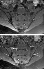
English: Magnetic resonance images of sacroiliac joints: psoriatic arthritis. Shown are T1-weighted semi-coronal magnetic resonance images through the sacroiliac joints (a) before and (b) after intravenous contrast injection. Enhancement is seen at the right sacroiliac joint (arrow), indicating active sacroiliitis. |
| 日期 | Published: 23 March 2006 |
| 来源 | Magnetic resonance imaging in psoriatic arthritis: a review of the literature. Arthritis Research & Therapy 2006, 8:207. doi:10.1186/ar1934 |
| 作者 | Fiona McQueen, Marissa Lassere and Mikkel Østergaard. |
许可协议[编辑]
| 注解 | 该图片含有注解:在维基媒体共享资源上查看注解 |
文件历史
点击某个日期/时间查看对应时刻的文件。
| 日期/时间 | 缩略图 | 大小 | 用户 | 备注 | |
|---|---|---|---|---|---|
| 当前 | 2009年2月6日 (五) 19:33 | | 1,004 × 1,586(1.07 MB) | Stevenfruitsmaak(留言 | 贡献) | {{Information |Description={{en|1=Magnetic resonance images of sacroiliac joints: psoriatic arthritis. Shown are T1-weighted semi-coronal magnetic resonance images through the sacroiliac joints (a) before and (b) after intravenous contrast injection. Enha |
您不可以覆盖此文件。
文件用途
全域文件用途
以下其他wiki使用此文件:
- ar.wikipedia.org上的用途
- az.wikipedia.org上的用途
- bs.wikipedia.org上的用途
- ca.wikipedia.org上的用途
- en.wikipedia.org上的用途
- es.wikipedia.org上的用途
- fa.wikipedia.org上的用途
- hi.wikipedia.org上的用途
- hu.wikipedia.org上的用途
- it.wikipedia.org上的用途
- outreach.wikimedia.org上的用途
- ta.wikipedia.org上的用途
- www.wikidata.org上的用途
- zh.wikipedia.org上的用途