File:Schizophrenia PET scan.jpg
Μετάβαση στην πλοήγηση
Πήδηση στην αναζήτηση
Δεν διατίθεται υψηλότερη ανάλυση.
Schizophrenia_PET_scan.jpg (224 × 248 εικονοστοιχεία, μέγεθος αρχείου: 23 KB, τύπος MIME: image/jpeg)
Λεζάντες
Λεζάντες
Δεν ορίστηκε λεζάντα
Σύνοψη
[επεξεργασία]
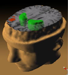
illustration of Schizophrenia's effect on the brain; taken from here
- Source: Andreas Meyer-Lindenberg, M.D., Ph.D., NIMH Clinical Brain Disorders Branch on the tree of life.
While patients performed a working memory task, the less the prefrontal cortex (red) activated, the more dopamine increased in the striatum (green).
Abstract of study is here.
Αδειοδότηση
[επεξεργασία]
This image is a work of the National Institutes of Health, part of the United States Department of Health and Human Services, taken or made as part of an employee's official duties. As a work of the U.S. federal government, the image is in the public domain.
 |
||
| Αυτό το αρχείο έχει χαρακτηριστεί ως απαλλαγμένο από γνωστούς περιορισμούς βάσει της νομοθεσίας περί πνευματικής ιδιοκτησίας, περιλαμβανομένων όλων των συναφών και συγγενικών δικαιωμάτων. | ||
Ιστορικό αρχείου
Πατήστε σε μια ημερομηνία/ώρα για να δείτε το αρχείο όπως εμφανιζόταν εκείνη την χρονική στιγμή.
| Ημερομηνία/Ώρα | Μικρογραφία | Διαστάσεις | Χρήστης | Σχόλιο | |
|---|---|---|---|---|---|
| τρέχον | 12:07, 30 Νοεμβρίου 2005 | | 224 × 248 (23 KB) | Skagedal (συζήτηση | Συνεισφορά) | illustration of Schizophrenia's effect on the brain; taken [http://www.nih.gov/news/pr/jan2002/nimh-28.htm from here] *Source: Andreas Meyer-Lindenberg, M.D., Ph.D., NIMH Clinical Brain Disorders Branch ''While patients performed a working memory t |
Δεν μπορείτε να αντικαταστήσετε αυτό το αρχείο.
Χρήση αρχείου
Δεν υπάρχουν σελίδες που χρησιμοποιούν αυτό το αρχείο.
Καθολική χρήση αρχείου
Τα ακόλουθα άλλα wiki χρησιμοποιούν αυτό το αρχείο:
- Χρήση σε ar.wikipedia.org
- Χρήση σε ast.wikipedia.org
- Χρήση σε bg.wikipedia.org
- Χρήση σε ca.wikipedia.org
- Χρήση σε cs.wikipedia.org
- Χρήση σε el.wikipedia.org
- Χρήση σε en.wikipedia.org
- Χρήση σε eo.wikipedia.org
- Χρήση σε es.wikipedia.org
- Χρήση σε fr.wikipedia.org
- Χρήση σε he.wikipedia.org
- Χρήση σε hi.wikipedia.org
- Χρήση σε hy.wikipedia.org
- Χρήση σε id.wikipedia.org
- Χρήση σε kn.wikipedia.org
- Χρήση σε mzn.wikipedia.org
- Χρήση σε no.wikipedia.org
- Χρήση σε pl.wikipedia.org
- Χρήση σε pt.wikipedia.org
- Χρήση σε ru.wikipedia.org
- Χρήση σε sk.wikipedia.org
- Χρήση σε sv.wikipedia.org
- Χρήση σε ta.wikipedia.org
- Χρήση σε tr.wikipedia.org
Δείτε περισσότερη καθολική χρήση αυτού του αρχείου.